Pancoast Tumor (Pneumonectomy)

This dissection is based on open procedures for right and left pneumonectomies.

Learning Objectives

Clinical Case: Lung Cancer

History & Physical and Labs
(Translation of shorthand is in italics)

CC: Left-sided shoulder and forearm pain.

HPI (history of present illness): AD is a 50-year-old man who describes a slow onset of pain over the course of two weeks. He reports no history of trauma. The pain is dull, aching and unremitting, and localizes to his left shoulder, along the medial border of the scapula and the medial side of the forearm. He reports dry skin on the left side of his face and left arm. He has not experienced fever, weight loss, headache, nausea, vomiting, dizziness, or bone pain.

PMH (past medical history): AD has with no significant medical or surgical history.

FH (family history):No history of malignancy in first-degree relatives.

SH (social history): AD is married with two grown children. He works for a general contractor whose primary business is to remodel old homes. He smokes one pack of cigarettes per week and drinks several beers per week. His wife also smokes.

Meds: None

Allergies: NKDA (no known drug allergies)


Physical Exam:

WN (well nourished), WD (well developed)

VS (vital signs): T=98.2 HR (heart rate)=68 RR (respiration rate)=15 BP (blood pressure)=140/90

HEENT (head, eyes, ears, nose, and throat): A ptosis of the left eye is evident. The left pupil is miotic (constricted) and accommodation is normal.

Neck: No JVD, no lymphadenopathy, strong carotid pulses, no bruits

Lungs: clear to auscultation on the right side; left side: decreased breath sounds and dullness to percussion near the apex

Heart: RRR (regular rate and rhythm), S1/S2 normal, no murmurs, rubs, or gallops ( = normal heart sounds, rate and rhythm)

Abdomen: moderately obese, soft, non-tender, non-distended, normal bowel sounds, no hepatosplenomegaly (hepato = liver; spleno = spleen; megaly = enlarged), no masses.

Extremities: Left sided diminished triceps reflex and weakness of finger adduction. Ulnar border of left arm and small finger tingles and is insensitive to soft touch.

Skin: Left upper limb and left side of face is dry and flushes with exertion.

Neuro: A&Ox3 (alert and oriented to person, time, and place), CN II to XII grossly intact. Skin is dry and flushes with exertion. Except as noted for the extremities, reflexes were normal and symmetric; strength, sensation normal.


Laboratory results: Lab tests were unremarkable.

Use the history & physical and laboratory data to answer the following questions:

(Click on the question to show the answer.)

AD reports shoulder pain especially along the medial border of the scapula. Which nerves serve this region?

From the upper thoracic spinal nerves, dorsal rami innervate muscle and skin along the vertebral column and ventral rami innervate intercosal muscles. The brachial plexus (ventral rami of C5 to C7) serves the shoulder. Cranial nerve XI serves the trapezius muscle, in the back and the sternocleidomastoid in the neck.

AD also reports pain on the ulnar side of his forearm and weakness is found in several arm muscles and reflexes. Which collection of nerves serve the arm?

The terminal branches of the brachial plexus. As you will appreciate when you study the brachial plexus, the distribution of nerve deficits indicates a problem with the ventral rami of C8 and T1.

In this patient, the dry skin and flushing of the skin upon exertion are caused by injury to a type of nerve. Which type of nerve regulates sweat glands and the diameter of blood vessels? Where would the affected nerves arise and which ganglia would be involved in the case of AD?

Sympathetic nerves of the autonomic nervous system. The preganglionic nerves would arise in upper thoracic levels. They would synapse in cervical or T1 paravertebral ganglia. The postganglionics would travel to the left side of the head and the left upper limb.

Which physical findings suggest a problem with the lungs? How could a lung tumor be responsible for the reported pain and nerve deficits?

On the left side, there are decreased breath sounds and dullness to percussion near the apex. The apex of the lungs extend superior to the clavicle. Therefore, a tumor in the apex would be in position to invade the body wall near the C8, T1 and T2 spinal nerves and the portion of sympathetic chain that supplies the head and upper limb.

Bonus question: Which elements of the social history are consistent with the possibility of lung cancer?

AD smokes and lives in a household of smokers. His work potentially exposes him to asbestos. Both are risk factors for lung cancer.

Diagnostic Studies:

Examine this CT of a Pancoast tumor. The red arrow shows the tumor invading a thoracic vertebra. The accompanying VH Dissector image shows a comparable axial slice. T3 is highlighted, but the tumor extends superiorly as high as T1. The spinal nerves, sympathetic chain, scapula, and first rib are included for reference. Rotate the image to demonstrate how the tumor would extend above the rib cage anteriorly.

Pancoast Tumor CT

To learn more about this mass, we order a percutaneous needle biopsy. Pathology Report: lung cancer.

If the cancer has spread to lymph nodes, the cancer has spread too far for a surgical procedure to be beneficial. Where should we look for lymph nodes? Why?

Lymphatics in the lungs drain to the mediastinum via the hilum of the lung. Therefore positive nodes would be in the mediastinum near intercostal spaces 2 and 3.

CT showed enlarged mediastinal nodes. To determine whether the nodes were positive for cancer, mediastinal nodes were sampled.

Pathology Report: inflammatory reaction; no evidence of cancer.

Decision: Resectable lung cancer.

Operative Approach

The operative approach for AD would attempt to preserve as much of the lung as possible. Later, we will consider the principles that underlie that attempt. Now, we break from the case and follow the procedures for completely removing the right and left lungs. Nowadays, this would be done thoracoscopically if possible. The following is based on a classical open procedure.

Click this link to set up your session. As you dissect, structures that have already been highlighted will be revealed. For clarity, the right arm and the pectoralis major and minor muscles have been removed. Mouse over the image with the dissection or highlight tool to identify the visible structures.

Right lung

  1. The incision is shown in blue for a lateral thoracotomy. The patient's arm is over their head, which rotates the scapula. The incision divides the rhomboid major, latissimus dorsi and serratus anterior.

    Incision for a Lateral Thoracotomy

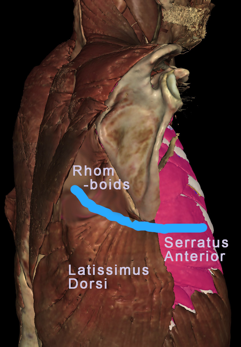
    To emulate this incision, dissect the latissimus dorsi and serratus anterior. (Note: the scapula is not rotated in VH Dissector; the above image was Photoshopped).

    What do the latissimus dorsi, rhomboids, and serratus anterior do?

    Principal functions: latissimus dorsi - adduct and extend the arm; rhomboids - medially rotate and retract the scapula (move it medially); serratus anterior - laterally rotate and protract the scapula (move it laterally).

  2. The rib cage and intercostal muscles are exposed. The external and internal intercostal muscle of the fourth intercostal space need to be divided from the ribs.

    Should you make your incision along the superior border of the rib or the inferior border? Why?

    An incision along the superior border avoids the neurovascular bundle traveling just inferior to each rib.

    Dissect the external oblique and then the internal oblique. Zoom in to see the intercostal nerve (yellow), vein (blue), and artery (red) peaking out below each rib.

  3. Typically, the surgeon would use retractors to spread open the intercostal space. For a better view remove ribs 3, 4, 5, and 6. This will give you a better view of the neurovascular bundle associated with each rib. Dissect the nerves, arteries and veins. The right lung is seen superiorly and the diaphragm inferiorly. Mouse over the lungs to identify the three lobes and find the borders between them. Use the highlight tool to de-highlight the middle lobe.

    To view the lungs, the surgeon has to divide the costal pleura creating a pnuemothorax. Why? What will happen to the lung?

    The lung will collapse, because 1) air pressure will equalize between the inside and outside of the lung, and 2) elastin fibers in the lung will contract. Think of the pleural membranes as a balloon. Imagine the lung as your fist and punch your fist into the balloon (i.e., inflate the lung) until the membrane covering your fist meets the membrane opposite it. To avoid compressing the air in the "balloon", you would remove air as you expand the lung. Normally, the lumen of the pleural membranes (the balloon) is a vacuum and air in the lungs keeps the visceral pleura of the lung pressed against the parietal pleura that lines the ribs, diaphragm, and mediastinum. When the rib cage expands and the diaphragm moves interiorly, space is created for the lungs to expand, and air is drawn into the lungs. Dividing the pleura allows air to enter and eliminate the vacuum. The lung will collapse to ~ 1/3rd it's normal size. The anesthesiologist can remove more air to make the lung smaller still, affording the surgeon lots of space.

  4. Dissect the middle lobe. Identify the bronchi and arteries of the middle lobe. Notice how arteries and bronchi identified by VHD branch together. If all the bronchi could be colored, each artery would have an accompanying bronchus. Be careful to preserve the lobar artery and bronchus, as you dissect all the branches labeled "segmental".

    What is the functional significance of bronchi branching together with arteries?

    As the airway and artery branch in parallel, they get narrower and narrower. When the airway is at its thinnest, so is the artery (now a capillary), and gases exchange between the two. The lung is divided into lobes and bronchopulmonary segments. The first branch of the trachea defines left vs. right lung. The second branch defines a lobe. The third branch defines a bronchopulmonary segment.

  5. Dissect the superior lobe. Now you see veins in addition to the arteries. Veins do not branch in parallel with bronchi. Instead, they travel between bronchopulmonary segments. Avoiding tbe veins, preserve branches labeled "lobar" as you dissect arterial and bronchial branches labeled "segmental".

  6. Dissect the lower lobe. Rotate the image to appreciate the distribution of airways, arteries, and veins. Leave the veins and dissect anything labeled "segmental".

  7. Observe the bronchopulmonary lymph nodes, colored green. Note that they are wrapped around the main and lobar bronchi. This region is the hialus of the lung (the region where vessels and airways enter/leave the lung). Dissect the lymph nodes.

  8. Trace the veins to where they enter the left atrium of the heart. The muscle of the left atrium (myocardium) is colored pink and the lumen of the atrium is brown. Dissect each vein. Rotate the image to find two rings of atrial myocardium. This where the veins entered the left atrium. Use the index to look up the right pulmonary veins. Toggle them on and off to confirm they enter the heart at this location. It will help to rotate the image as you do this.

  9. By now, your dissection should look something like this. For clarity, we highlighed the superior vena cava and de-highlighted the superior lobar artery. This view looks head-on at the hilum of the right lung. The hilum is bounded by the superior vena cava, arch of the azygos vein and azygos vein. Mouse around to identify the: pulmonary artery; superior, middle, and inferior lobar arteries; trachea; main (primary) bronchus; superior, middle, and inferior lobar (secondary) bronchi . Determine which primary branch is most anterior. The pulmonary veins have been removed from this view of the hilum. To confirm they lie inferior to the airway and artery, infer their position by re-identifing the left atrium, as viewed through anular rings of myocardium.

    Which regions are drained by the arch of the azygos vein?

    The posterior walls of the chest and abdomen.

    Bonus question: In what order do you imagine a surgeon would divide the structures within the hilum?

    Historically, the surgeon divided the artery first, because it is anterior most and exposes the main bronchus. After dividing the bronchus, the pulmonary veins are divided. The logic was that if you divide the veins first, the arterial pressure would increase too much. Although true for removing other organs, pulmonary arterial pressure is low to begin with. Consequently, the increased blood pressure is not a big factor. Current data indicates that cancer is more likely to spread if you divide the artery first. Consequently, surgeons now start with the pulmonary veins and work their way superiorly.

  10. Recall that for artery and bronchi there is one primary branch for each lung and one secondary branch for each lobe of the lung. The next, tertiary branches, define functional subunits of each lobe, known as bronchopulmonary segments.

    How would you use this information to perform a partial pneumonectomy?

    Instead of dividing the main bronchus and pulmonary artery, you would divide the lobar branches for a lobectomy, or the first segmental (tertiary) branche(s) to remove one (or more) bronchopulmonary segments. By re-inflating a tertiary bronchus, that bronchopulmonary segment is clearly distinguished from the rest of the lobe. This enables removal of the lung without compromising neighboring bronchopulmonary segments

  11. Identify two additional structures near the hilum that might be injured during a surgical procedure to divide the right hilum and its contents: the vagus nerve and the phrenic nerve (the accompanying phrenic artery and vein were not identified by VHD).

    What is the consequence of injuring the right vagus nerve?

    Parasympathetic innervation would be compromised for the embryonic, right side of the esophagus and abdominal 2/3 of the GI tract.

    What is the consequence of injuring the right phrenic nerve?

    Paralysis of the right hemi-diaphragm would impair breathing.

  12. Rotate the image to appreciate the three-dimensional relationships. End up with this view to examine the hilum along its edge.

Left lung

The left lateral approach to lung is similar to right lateral approach. The beginning of this dissection corresponds to step (4), above.

  1. Click this link to set up your session. Note that we also removed ribs 7 and 8 for clarity.

  2. Use your mouse to identify the upper and lower lobes of the left lung. De-highlight the lower lobe to appreciate the boundary between the two. Rotate the image to find the superior-most pole of the lower lobe. Dissect the scapula and the superior serratus posterior. This will allow you to identify vertebrae.

    Which vertebra corresponds to the superior-most pole of the lower lobe?

    T4. This is also true on the right side. To auscultate the superior lobe from the back, your stethoscope needs to be placed above T4. Otherwise you are listening to the lower lobe.

    Dissect the lower lobe.

  3. Identify (but do not memorize!) all the segmental arteries and trace them proximally to the left pulmonary artery (dark brown because it is not highlighted). Note that there is no lobar artery. Dissect the segmental arteries. Preserving the left superior lobar bronchus, repeat this procedure for the segmental bronchi. Rotate the image until you see the inferior pulmonary vein enter the left atrium of the heart.

  4. Rotate the image to an anterior view. Notice how the inferior aspect of the heart is not covered by the left lung. This is the cardiac notch and is where a needle can be used to enter the pericardial sac without puncturing the lung. Dissect the left lung to view more of the heart. Use control-(or command-)z to replace the lung. Toggle the lung on and off to appreciate the thin portion of lung that covers the anterior surface of the heart. With the lung dissected, rotate the image to a lateral view to appreciate the thin space between the heart and the anterior chest wall. The portion of the lung that occupies this space is the lingula (tongue-shaped). The lingula corresponds to the middle lobe of the right lung. It is smaller than the right middle lobe, because the heart occupies the space where the middle lobe would be.

  5. Identify and dissect the superior and inferior lingual arteries, and then the lingual artery. Trace all the segmental arteries back to the pulmonary trunk and dissect them. Preserving the left inferior and superior lobar bronchi, repeat this procedure for the segmental and lingual bronchi. Rotate the image until you can see both lobar bronchi branching from the left main bronchus. Dissect the lobar bronchi.

  6. Rotate back to a lateral view. Trace the pulmonary veins to where they enter the left atrium. Dissect the pulmonary veins and highlight the left pulmonary artery. By now, your dissection should look something like this. Instead of the arch of the azygos vein, the left hilum of the lung is related to the arch of the aorta. As with the right lung, a careless surgeon might injure the left phrenic or left vagus nerve. Note the branch of the vagus nerve, the left laryngeal nerve. Not seen are cardiophrenic nerves that branch from the left vagus nerve at this point.

  7. Rotate the image to appreciate the three-dimensional relationships. End up with this view to examine the hilum along its edge.

  8. Compare this axial section through the left and right hila to this analogous slice of a CT.

    Pulmonary Artery CT

    The slices are not exactly the same. Use the arrow keys to move the VHD image superiorly and inferiorly. Be sure you mouse is hovered over the Axial window. Scan the VHP image to find the branch point (Carina) of the trachea and the inferior pulmonary artery entering the left atrium.

Take Home Lessons

When performing this procedure a surgeon should be aware of the following important anatomical concepts: