Hip Bone
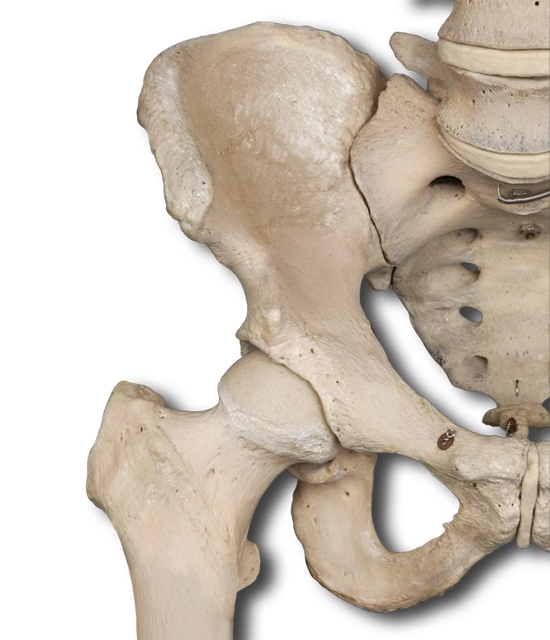


As you can see, the pelvic girdle is composed of two hip bones, also known as coxal bones, that unite anteriorly at the pubic symphysis and posteriorly with the sacrum at the sacroiliac joints (we'll look at that joint in a moment). The pelvic girdle supports the weight of the upper body, allows lower limb movement via the hip joint, provides muscle attachment points, and supports and protects abdominopelvic organs.
The bony pelvis is the complete ring of bones that forms a deep, basin-like structure. Identify the following:
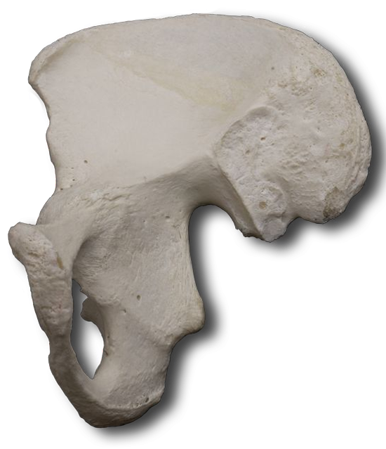
A newborn's hip bone is actually three separate bones that will ultimately fuse together. These three bones are not identified in your specimen, so use the interactive image to identify the following:
The ilium is the largest of the three coxal bones. Continue using the interactive images as a guide, identify the following on your specimen.
The ischium is the inferior, posterior portion of the hip bone. Using the same image as a guide, identify the ischium and the following ischial markings.
The pubis is the anterior and inferior part of the hip bone. Use the intereactive image to help identify which part of the coxa this is on your specimen.
Rotate your specimen to 45° and 90° to best observe the following structures. The pubis and the ischium surround the obturator foramen, the largest foramen in the body. It decreases the weight of the coxa. The fibrous obturator membrane (not shown) covers the foramen, except one small area where blood vessels and nerves pass between it and the bone.
The acetabulum is the deep fossa formed by all three bones. It is the socket that accepts the rounded femur head to form the hip (coxal) joint.
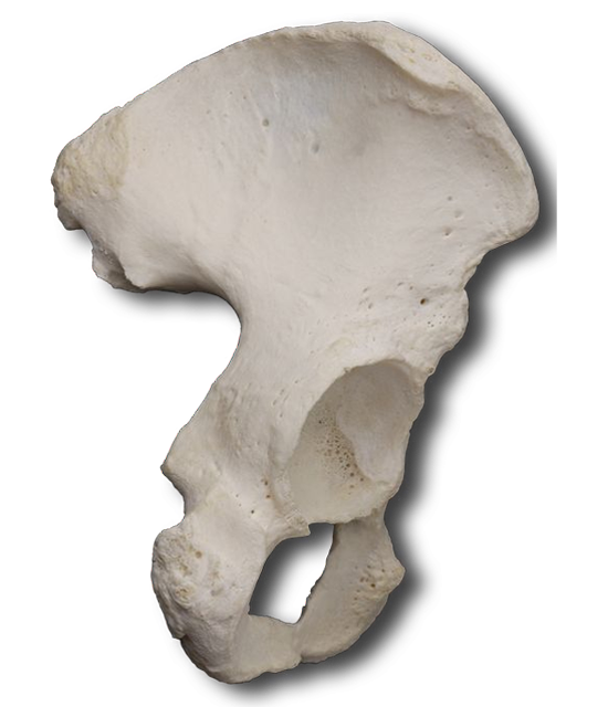
Note the female pelvis is wider and the pelvic inlet and outlet is larger. Additionally, the female pelvis has a subpubic angle greater than 90° (angle formed below and between the two pubic bones). These differences are child birth adaptations.
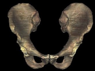
Male Pelvis
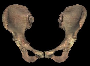
Female Pelvis
This is the final segment of your journey down ossification lane! Since you've already mastered the bones of the upper extremity, the very analogous structures here in the lower half of the body should be much easier to learn.
Identify the following:
You may notice a different use of terminology within the title of this study. You probably refer to everything below your hip as being "the leg." Technically, this is not the case. Your thigh is the proximal portion of your lower limb. The "leg" is between the knee and the proximal foot.
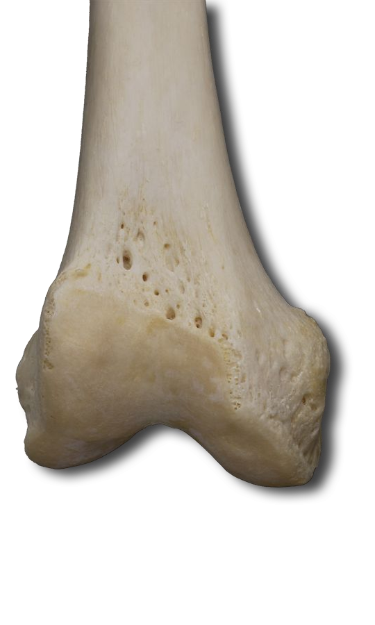
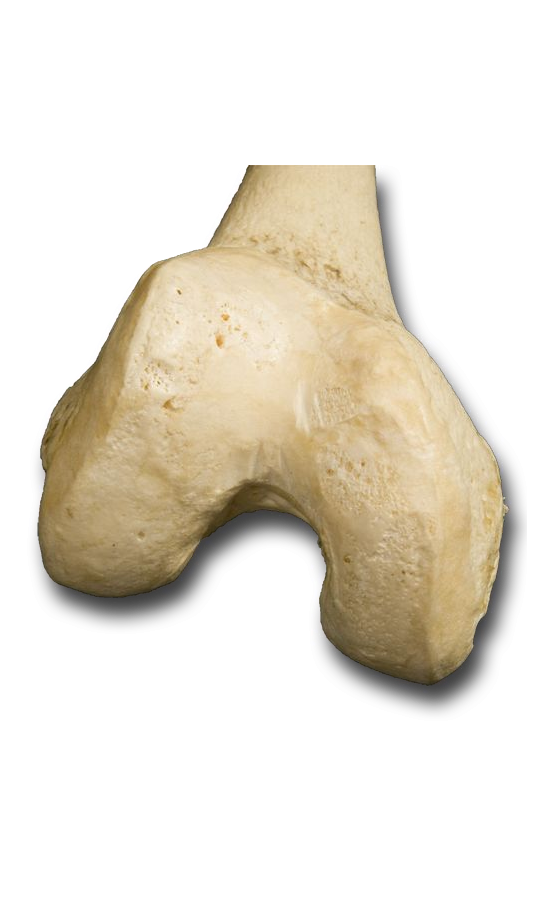
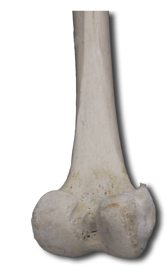
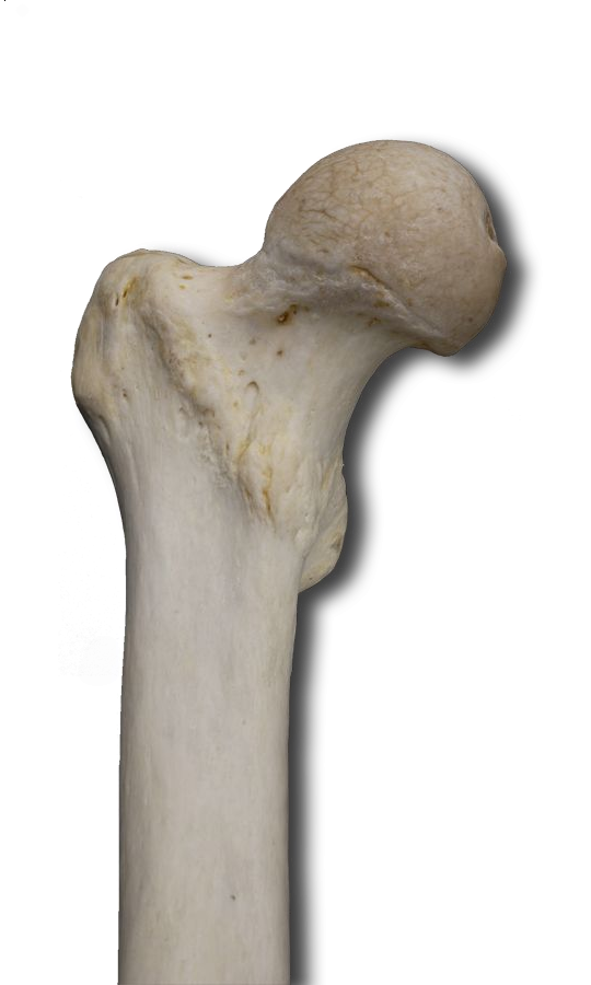
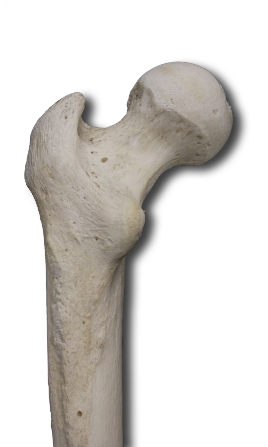
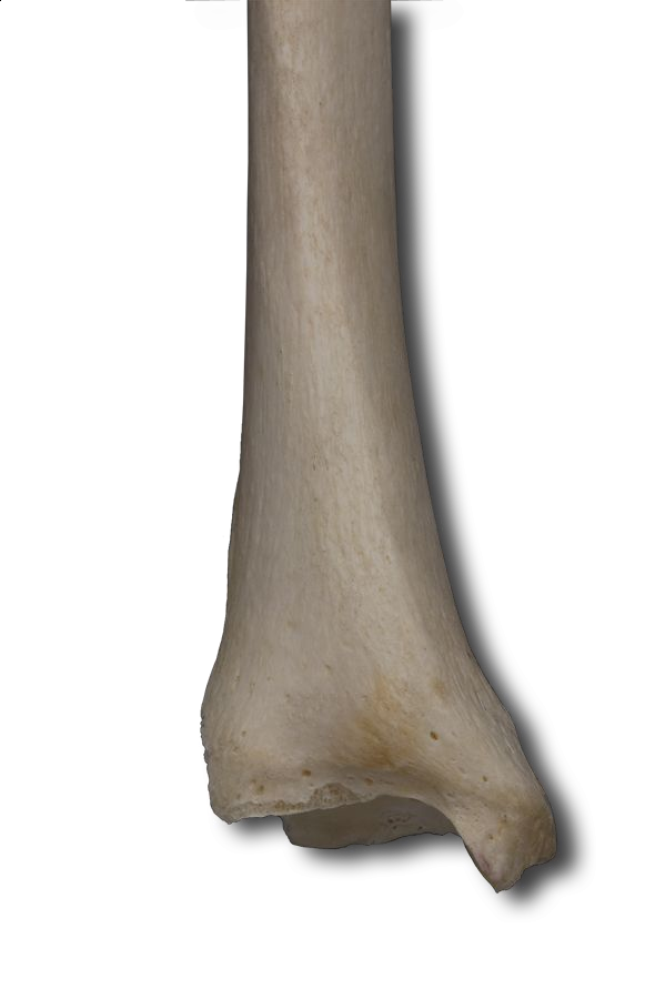
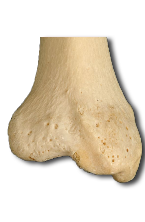
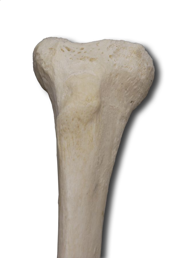
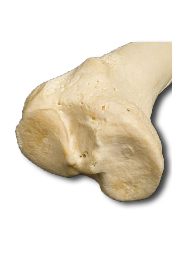
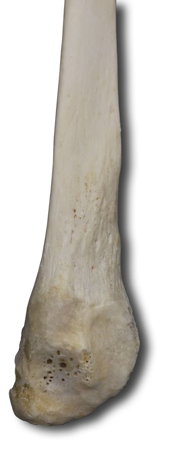
The femur is the single thigh bone. It is the largest, longest, strongest bone in the body. Its durable anatomy reflects the fact that the stress on the femur during vigorous jumping can reach 280 kg per square centimeter. This is about 2 tons per square inch!
Using the interactive image as a guide, identify the following femoral markings in your specimen, and record them on your worksheet. As always, rotate and manipulate your specimen for the best viewing perspective.
This sesamoid bone develops in the tendon of the quadriceps femoris muscle. It protects the tendon from damage and increases the force that the muscle applies to the leg.


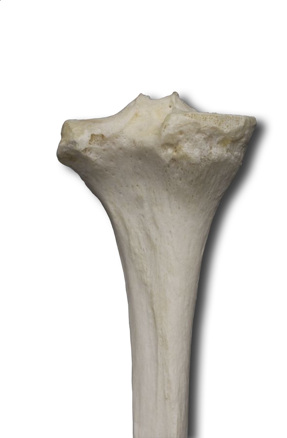


You have already identified these two bones, however, do so again to reinforce the fundamental anatomy. Using the interactive image as a guide, identify the following tibial and fibular markings.
The seven ankle bones transfer weight from the tibia to the metatarsals and, in the case of the calcaneus, actually supports body weight. When we step forward to our heel, we're placing our entire body weight on the calcaneus. Let's identify all seven of these bones. Your specimen is initially turned to 45° for a good perspective of the anterior right foot and the medial left foot. This is simply due to the general posturing of the specimen. Use the rotation wheel to gain the best perspective as you identify the tarsals and metatarsals.
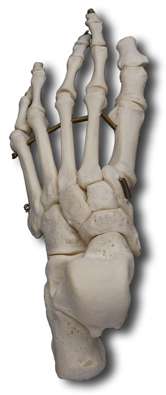


MILC No Thanks Cow - That little pneumonic should help your memory starting with the distal row moving medial to lateral then the proximal row moving medial to lateral once again. Identify each of these on your specimen.
Note that during walking the talus transmits some body weight to the metatarsals through the calcaneus and cuboid bones (and the ground when the calcaneus is in contact with it). The remainder of the body weight on the talus is transmitted through the navicular and three cuneiform bones to the metatarsals.
Identify the metatarsals on the interactive image. These five bones make up the intermediate portion of the foot which transfers weight to the ball of the foot and the phalanges. These bones are simply numbered. Identify each in your specimen.
Identify the phalanges on the interactive image. For those Friends fans in the class, remember Phoebe's alter-ego, "Regina Phalanges"? No real point here, other than reaching for a way to remember the terminology! :-) As you see the foot phalanges follow a pattern VERY similar to that evolved in the hand. Only our great toe (big toe) has two phalanges. The other four toes have three, proximal, medial, and distal.
Now that you are "hip" to the lower extremities, we can place a proverbial (knee) cap on the skeletal system and move right on with the conclusion of this system. The next time you hear "the leg bone's connected to the knee bone", I expect each of you to clarify this is referring to the patella's articulation with the tibial tuberosity. :-) Isn't it amazing how science can take the fun out of everything!