Bony Thorax
Directions
- Print this PDF worksheet for a hardcopy guide as you work through this lesson.
- Within the lesson click the red linked headings to bring up the desired starting point within the cadaver for your work.
- Use the provided images on the worksheet to annotate and identify specific anatomical structures.
The thoracic cage is the bony enclosure formed by the sternum, costal cartilage, ribs, and bodies of the thoracic vertebrae. It encloses and protects the thoracic organs and provides muscle attachment points.
This is the midline/anterior part of the thoracic cage. Three sternal bones become fused together in the elderly. It is the attachment point for chest and neck muscles responsible for moving the arms and head. It also protects internal organs particularly the heart. Locate each of the following in your specimen:
- manubrium - This is where the clavicles attach.
- body - The middle and largest sternal bone. It articulates directly or indirectly with the costal cartilage of the second through tenth ribs.
- sternal angle - This is the joint between the manubrium and the body. It marks the level at which the cartilage of the second rib joins the sternum. This joint is clearly visible in your specimen, but not specifically identified.
- xiphoid process - This is an attachment site for some abdominal muscles. It is hyaline cartilage during infancy and childhood and doesn't completely ossify until about age 40.
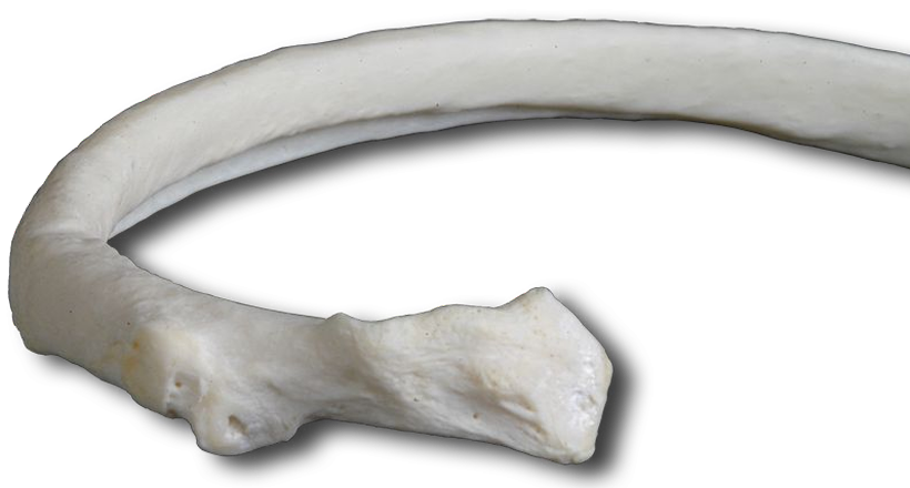
The 12 rib pairs provide structural support to the sides of the thoracic cavity. As you can see, they increase in length from the first through seventh then decrease in length to the twelfth. Each rib articulates with its corresponding thoracic vertebrae. Notice how costal cartilage attaches MOST ribs to the sternum. Rotate your specimen to the anterolateral position for a better view.
- true (vertebrosternal) ribs - Ribs 1-7 attach directly to the sternum via costal cartilage.
- false (vertebrochondral) ribs - Ribs 8-10 attach to each other via costal cartilage and then to the cartilage of the seventh rib.
- floating ribs - Ribs 11 and 12 lack any costal cartilage connection to the sternum. The floating ribs are also "false ribs" but not referred to as vertebrochondral.
The ribs allow the thoracic cage to change shape and size during respiration.
Use the interactive image to identify the:
Note how the ribs articulate with the vertebrae at both the facet and the tubercle.
Self-test Labeling Exercises