Cranial Bones
Directions
- Print this PDF worksheet for a hardcopy guide as you work through this lesson.
- Within the lesson click the red linked headings to bring up the desired starting point within the cadaver for your work.
- Use the provided images on the worksheet to annotate and identify specific anatomical structures.
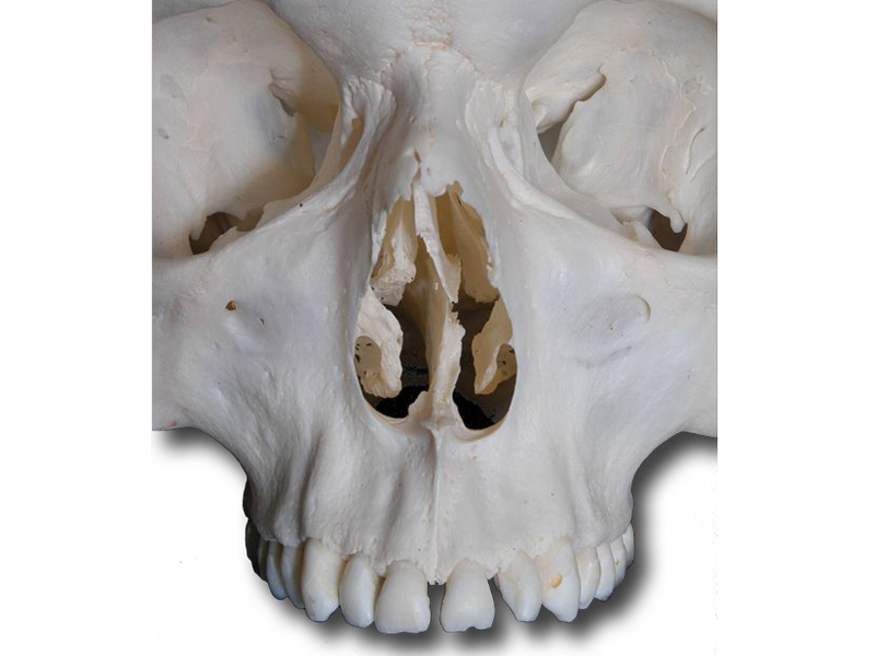
Using the highlight tool identify the following bones of the anterior skull.
- frontal bone
- zygomatic bones (right & left)
- maxilla
- mandible (note: 9 parts of the mandible are identified. At this point you are only responsible for the single unified bone.)
- nasal bones (right & left) (note: these are identified as a single bone in your virtual cadaver)
- ethmoid
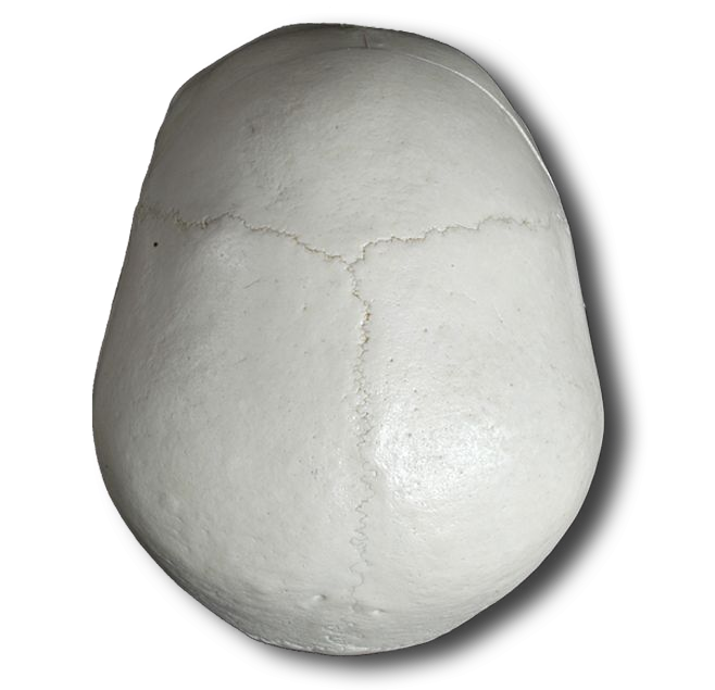
At birth, the frontal bone has two parts separated by a frontal suture (immovable joint). This suture generally disappears by the age of six when the frontal bones unite.
Identify the bones and test your identification knowledge with your virtual cadaver. Additionally, you should mark the diagrams in your worksheet. Labeling and highlighting diagrams is VERY helpful when learning anatomy.
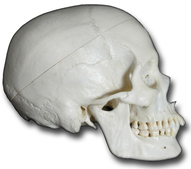
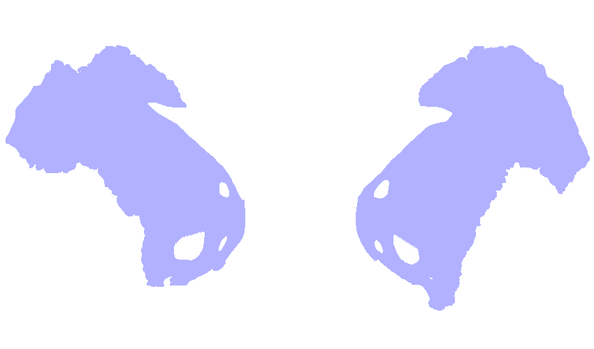
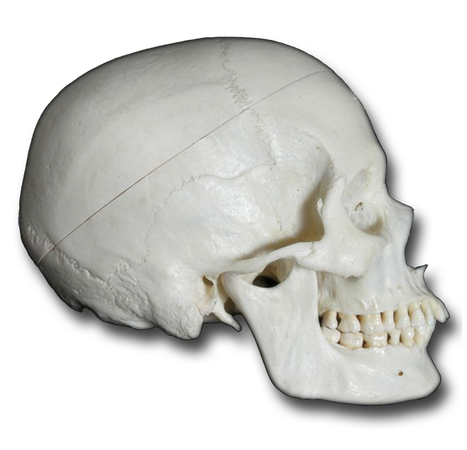
Again, using the highlight tool identify the following bones of the lateral skull.
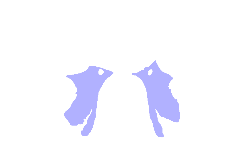
- temporal bone (right)
- frontal bone
- parietal bone (right)
- occipital bone (note: 2 parts of the occipital bone are identified. You, however, are only responsible for the single unified bone at this point.)
- maxilla
- mandible
- zygomatic bone (right)
- sphenoidal bone
- nasal bone (right)
Using the interactive image, identify the following and record them on your worksheet.
- supraorbital margin - This thickened brow of bone provides protection for the eyes.
- supraorbital foramen - Foramena are bone openings. This small opening is located in each supraorbital margin. Nerves and blood vessels pass through these openings to supply the forehead and scalp.
Again, using the interactive image, identify the following parts of the temporal bone.
- squamous portion - This simply forms part of the protective brain case.
- mastoid portion - Again, forms part of the protective brain case.
- zygomatic process - Together with the temporal process of the zygomatic bone, this forms the zygomatic arch, the "cheek bone." It is the attachment point for a muscle moving the mandible and for ligaments holding the mandible in place.
- external auditory meatus - the opening through which sound waves are conducted to the ear
- mastoid process - the rounded projection that is the attachment point for several neck muscles
- styloid process - This process is clearly evident in your specimen and identified by the software with a simple "mouse over". This is the attachment point for muscles and ligaments of the tongue and neck.
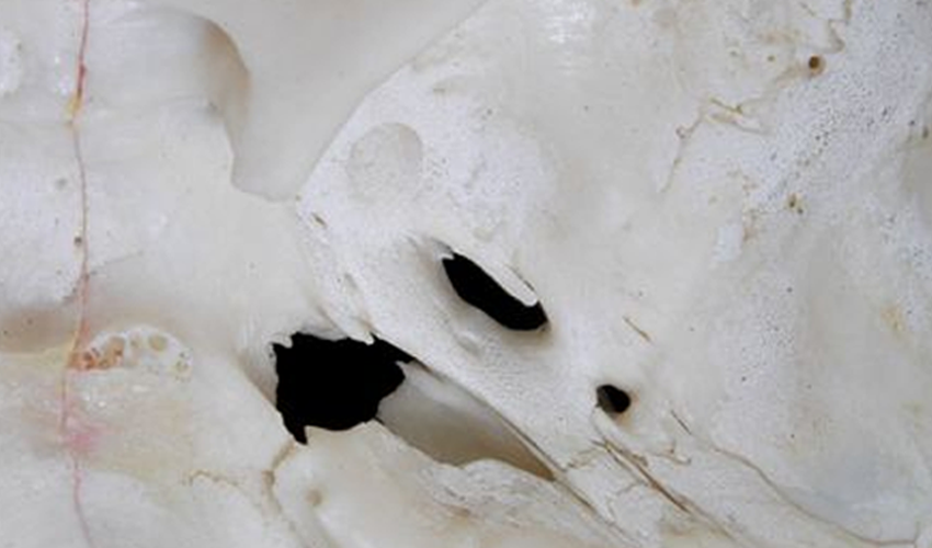
- stylomastoid foramen - Located between the styloid and mastoid processes, this opening allows the facial nerve (shown in yellow) to leave the skull.
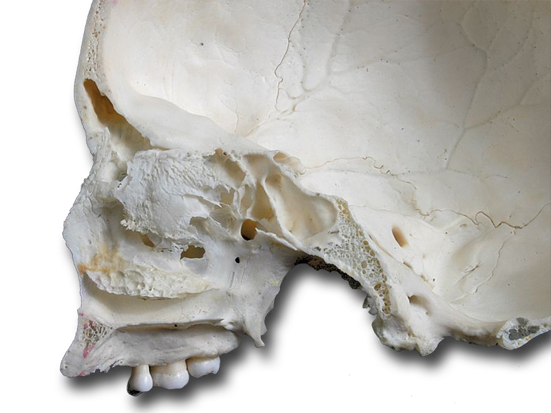
Dissect away the maxilla, frontal, and nasal bones.
Locate and highlight the ethmoid. Ethmoid means "like a sieve" which is a bit apparent from your dissection. Structurally, the ethmoid forms part of the cranial floor, the medial wall of the orbits (eye sockets), part of the nasal septum, and part of the sidewalls of the nasal cavity.
Using the interactive image, identify each of the following parts of the ethmoid, which are too small to observe in your dissection.
- perpendicular plate - This part of the ethmoid forms part of the nasal septum.
- cribriform plate - This part of the ethmoid forms the roof of the nasal cavity.
- olfactory foramina - These are small openings in the cribriform plate that pass olfactory nerves to the nasal cavity for the sense of smell.
- crista galli - This midline projection of the ethmoid is the attachment point for a membrane holding the brain in place.
- inferior and middle nasal conchae - These scroll shaped projections of the ethmoid increase the surface area of the nasal cavity.
Dissect away the maxilla, the right and left zygomatic bones, the ethmoid, and the nasal bone.
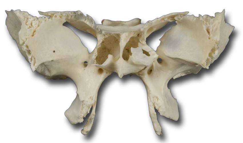
Sphenoid means "wedge shaped" but from the anterior view it looks more like a butterfly. It is frequently referred to as the "keystone of the cranial floor" because it articulates with all the other cranial bones and holds them together. From a superior vantage point, you can really see how it is central in its location and holds the surrounding cranial bones in place.
Using the interactive image, identify each of the following parts of the sphenoid. As with the ethmoid, these structures are not easily viewed in the dissection.
- body - the cube-like medial portion between the ethmoid and occipital bones
- sella turcica - This bony, saddle-shaped structure on the superior surface of the body forms the space containing the pituitary gland.
- greater wings - These lateral projections from the body form part of the lateral wall of the skull.
- optic foramen (canal) - This is the opening through which the optic nerve passes providing the sense of vision.
- pterygoid processes - These are prominent projections off the inferior sphenoid. Click the "Index" tab and type "medial pterygoid" into the search box. Hold the shift key down and click both the right and left medial pterygoid muscles then click the "Add & Highlight" button. As you can see, the pterygoid processes are attachment sites for muscles and ligaments of the mandible (jaw) and for some throat muscles.
- foramen ovale - The mandibular nerve passes through this opening.
- foramen lacerum - This opening is partly covered by a fibrocartilage layer with a few blood vessels passing through to supply the dura mater (a membrane covering the brain). The carotid canal opens into the superior part of the foramen.
- foramen rotundum - This opening provides passage for the maxillary nerve which subdivides to form the superior alveolar nerves serving the upper teeth.
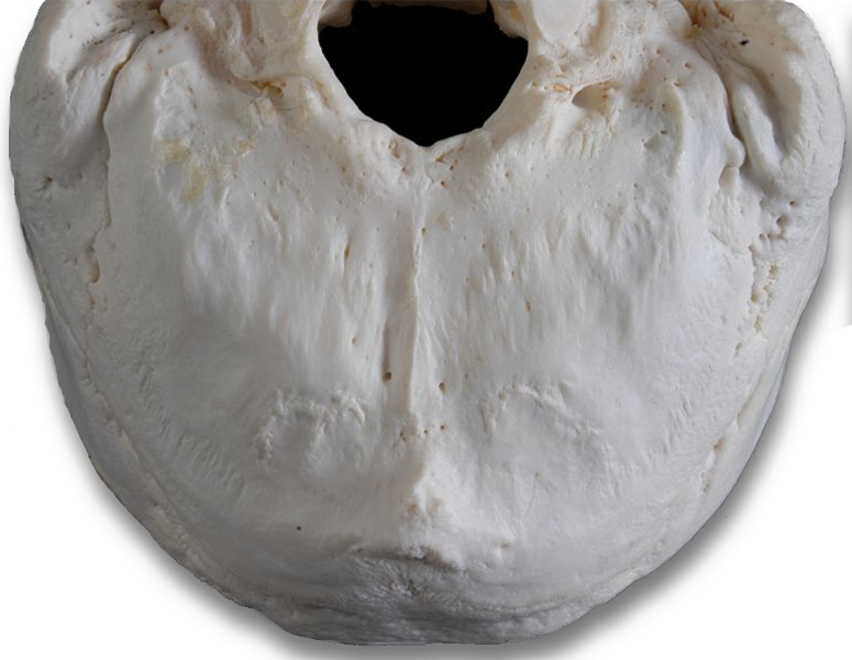
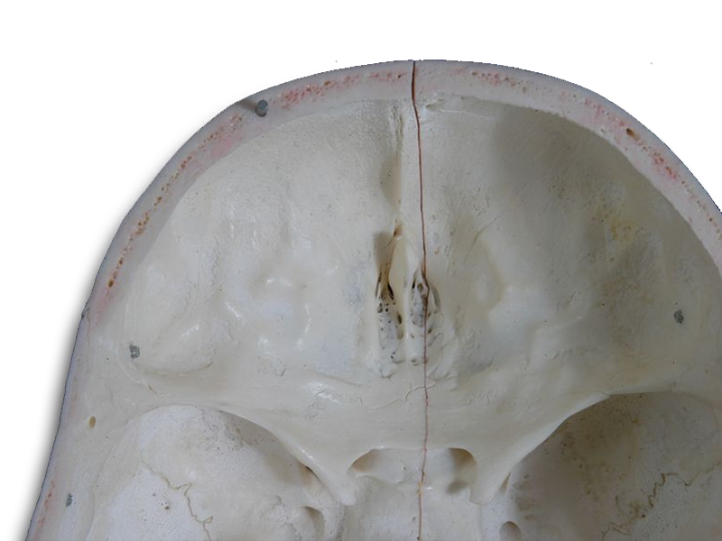
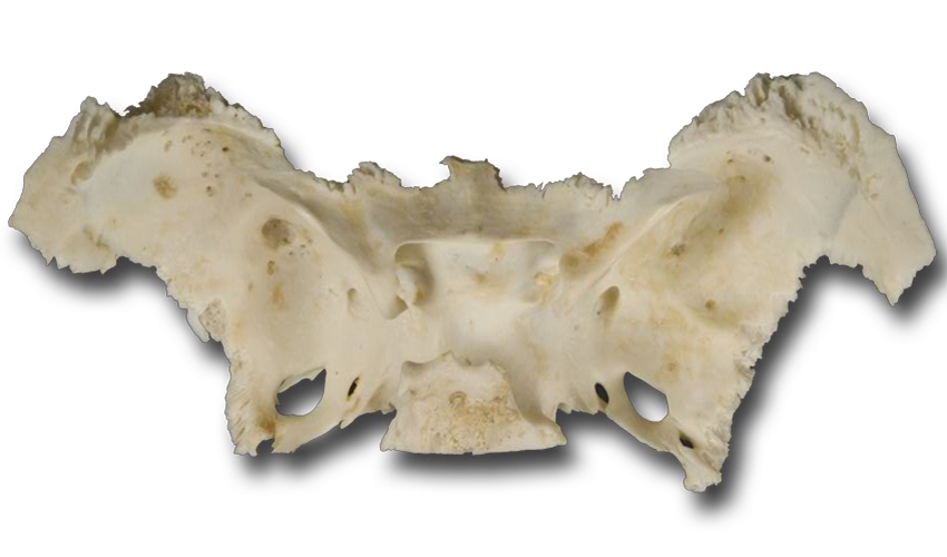
Using the highlight tool identify the following bones of the posterior skull.
- occipital bone
- temporal bones (right & left)
- parietal bones (right & left)
- sphenoidal bone
Locate the following with the interactive image. Then locate them on the cadaver, and record them on your worksheet.
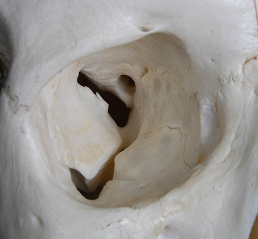
- foramen magnum - The inferior part of the brain, the medulla oblongata, connects with the spinal cord within this opening. Vertebral and spinal arteries also pass through it.
- occipital condyles - These two oval processes on either side of the foramen magnum have convex surfaces. They articulate with depressions on the first cervical vertebra (atlas) which forms the atlanto-occipital joint. Joint movement allows us to tilt our head up and down as you nod "yes".
- external occipital protuberance - This prominent projection is where the ligamentum nuchae, a large fibroelastic ligament, attaches to help support the head. This ligament extends to the seventh cervical vertebrae. Other muscles that move the skull also attach here.
- superior and inferior nuchal lines - These raised ridges on the occipital bone are attachment sites for skull moving muscles.
Sutures are the immovable joints between skull bones. Names, such as the frontozygomatic or sphenoparietal sutures, clearly reflect the bones they unite. Others, such as the coronal suture, do not.
These sutures are clearly visible in your dissection, but they are not specifically identified. Identify each of the following. Then locate them on your specimen, and record them on your worksheet.
Sutural bones, also called wormian bones, are small bones located within the sutures. The number of sutural bones varies widely between individuals.
Self-test Labeling Exercises