Vertebral Markings


The vertebral column consists of 26 bones with the following functions:
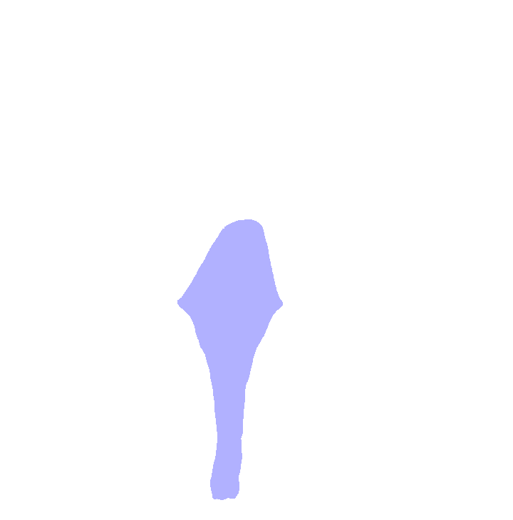
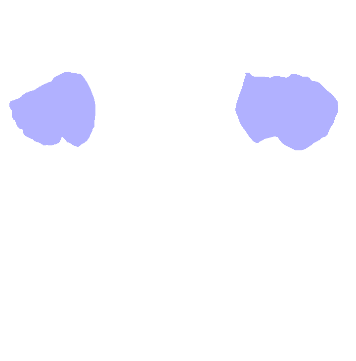
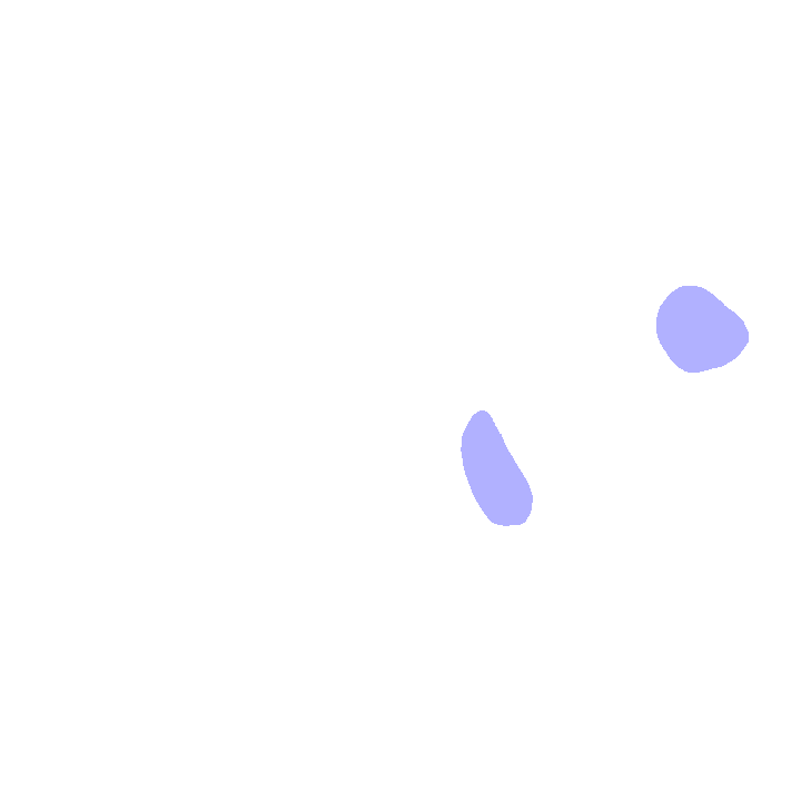
Identify the following:
When do you eat your meals? Seven, twelve, and five are pretty common answers AND that's the number of cervical, thoracic and lumbar vertebrae we possess! :-)
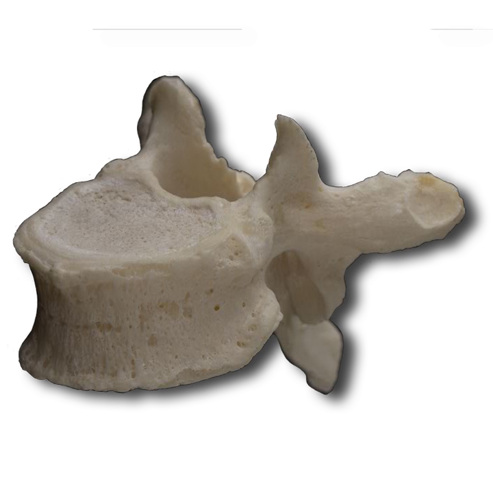
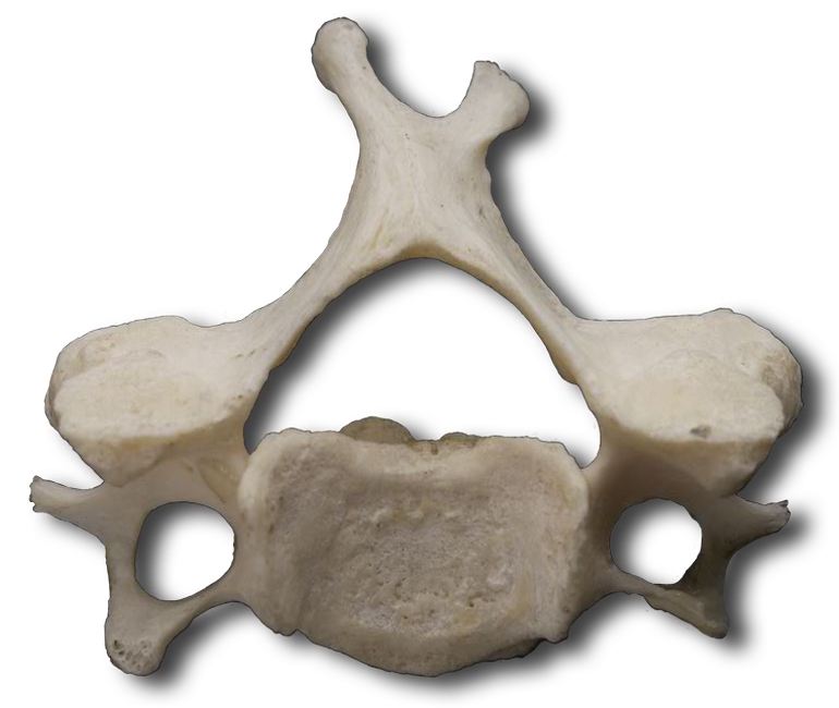
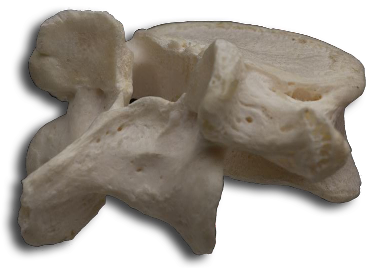
We'll use C3 to identify some general vertebral markings and characteristics. You'll soon discover why we don't use the atlas or axis for this purpose since they differ significantly from the others.
Locate the following with the interactive image and correlate anatomy to the specimen. Identify each on your worksheet:
Label these normal curves on your worksheet. Newborns have a single anteriorly concave curve throughout the length of their vertebral column. The cervical curve develops at about 3 months as the head begins to be held erect. Later, as the child sits up, stands, and walks, the lumbar curve develops. The thoracic and sacral curves retain their original curvature and are called "primary curves." Cervical and lumbar curves are called "secondary curves" since they develop after birth.
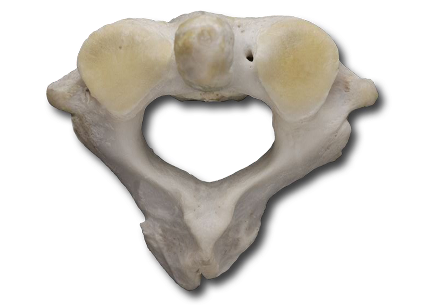
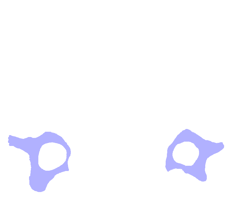
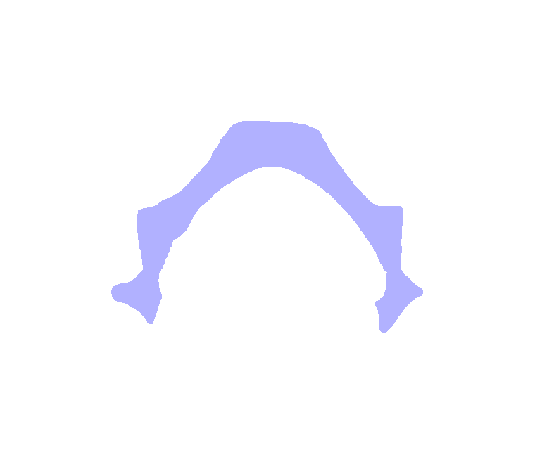
Note that cervical vertebrae are smaller than thoracic or lumbar vertebrae. All cervical vertebrae have 3 foramina, 1 vertebral foramen and 2 transverse foramina through which the vertebral artery and its accompanying vein and nerve pass. The spinous processes of C2 through C6 are often split into two parts (bifid). Using the following interactive images to help, locate these foramina on your specimen and label them on your worksheet.
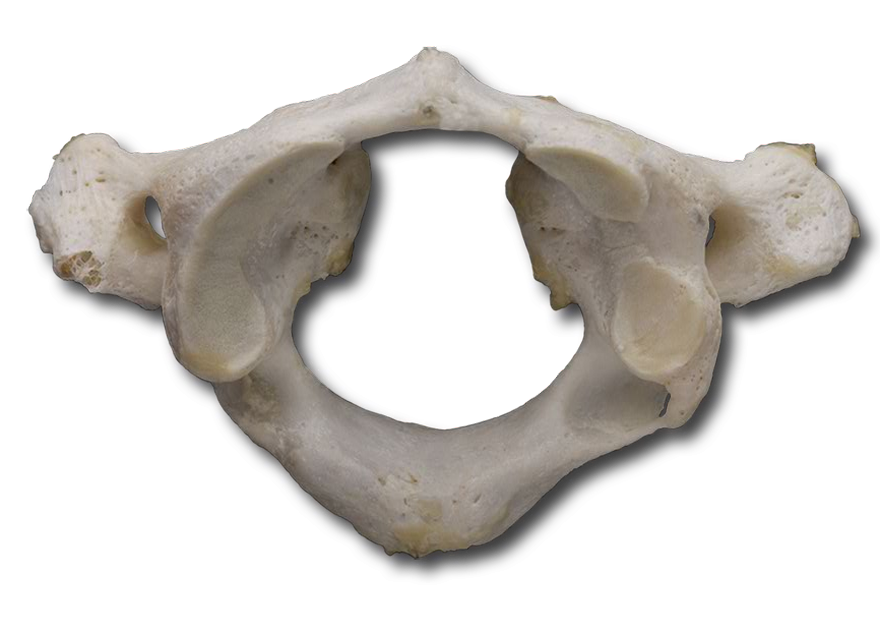
Named for the mythical person Atlas who supported the world on his shoulders, the atlas (C1) is a ring of bone upon which the head rests and moves. Using the image below, identify each of the following in your dissection and label them on your worksheet. Rotate the dissection as needed to obtain views from different angles:
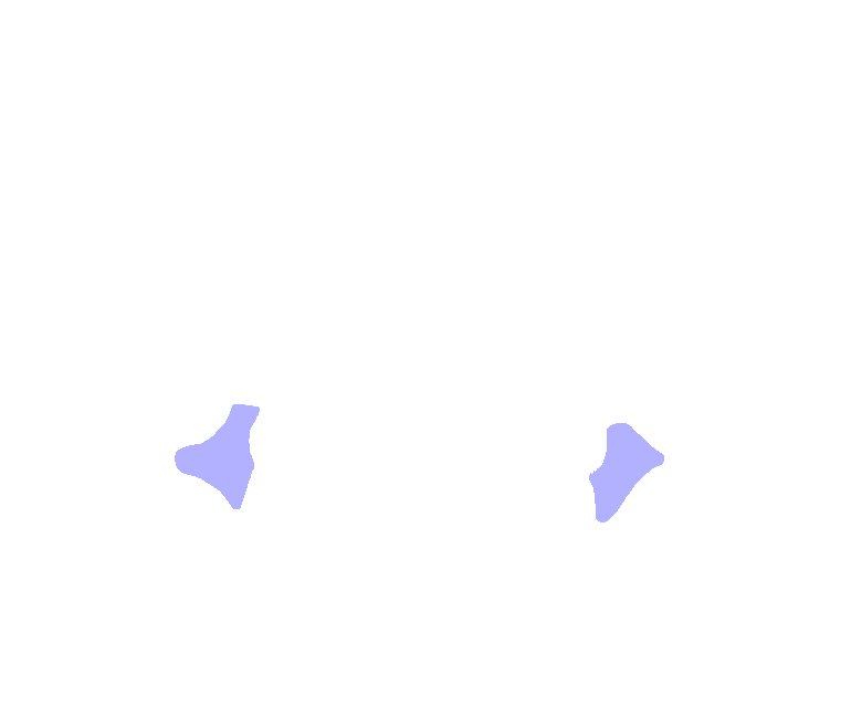
The following structures are on the Atlas only.
The following structures are visible on both the Atlas and the Axis.
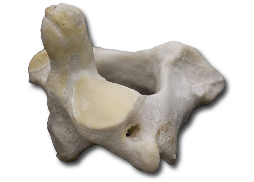
Dissect away the atlas (C1) to isolate the axis (C2) cervical vertebra.
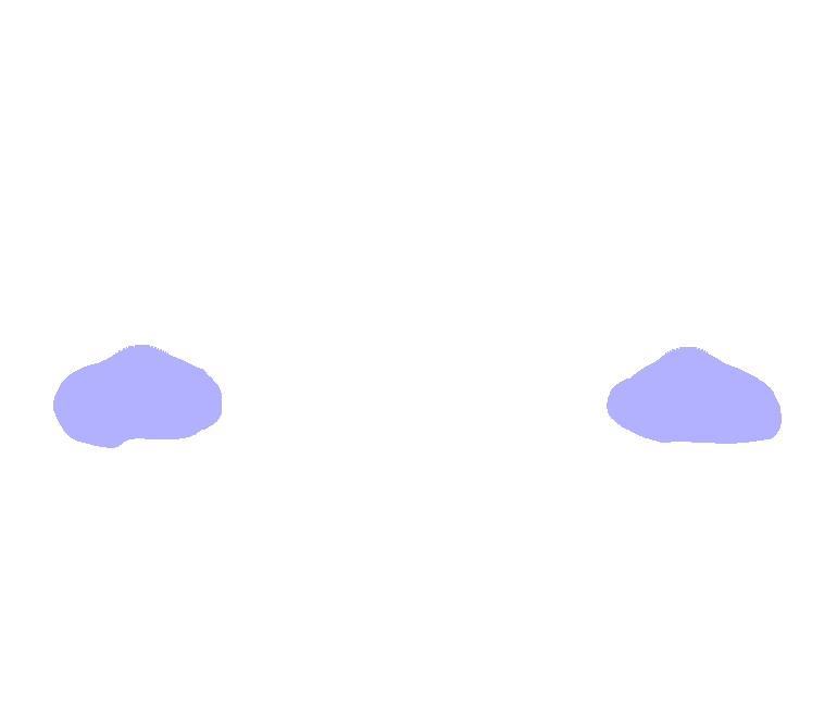
The following structure is on the Axis only.
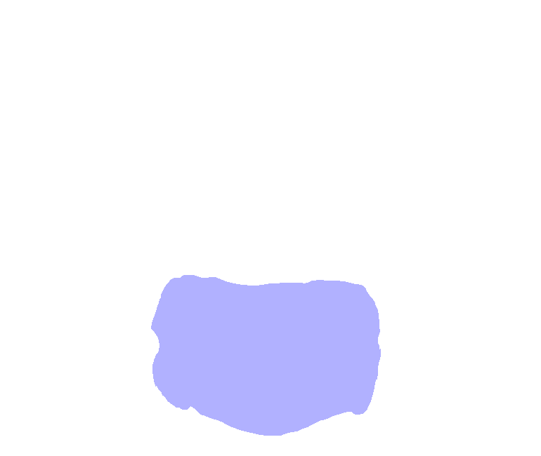
C3-C6 are typically structured vertebrae which were previously examined. C7, vertebra prominens, however, is somewhat different. Rotate your specimen for a lateral view, and identify the large spinous process. This process can be seen and felt at the base of the neck.
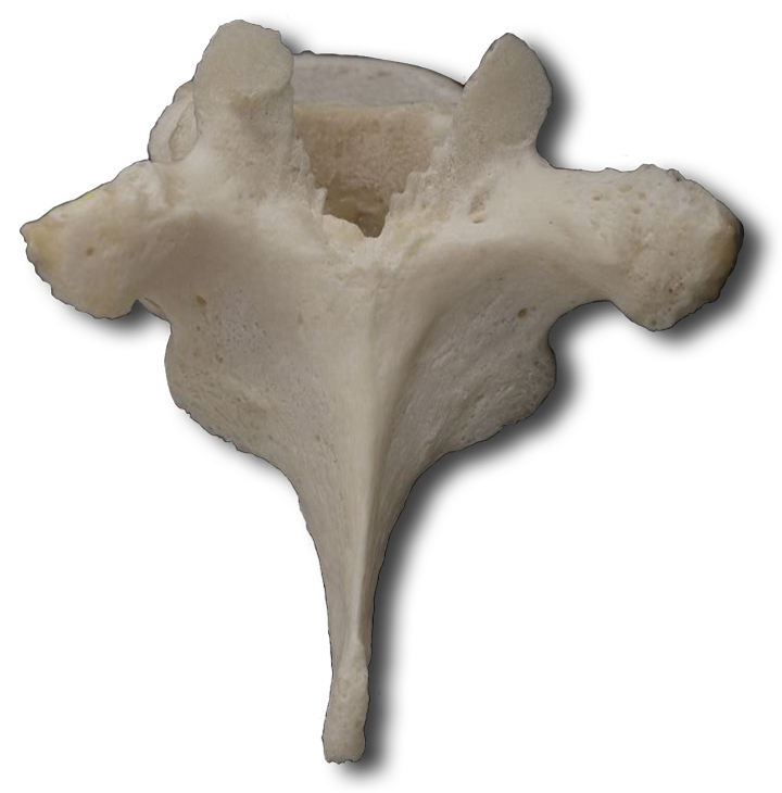
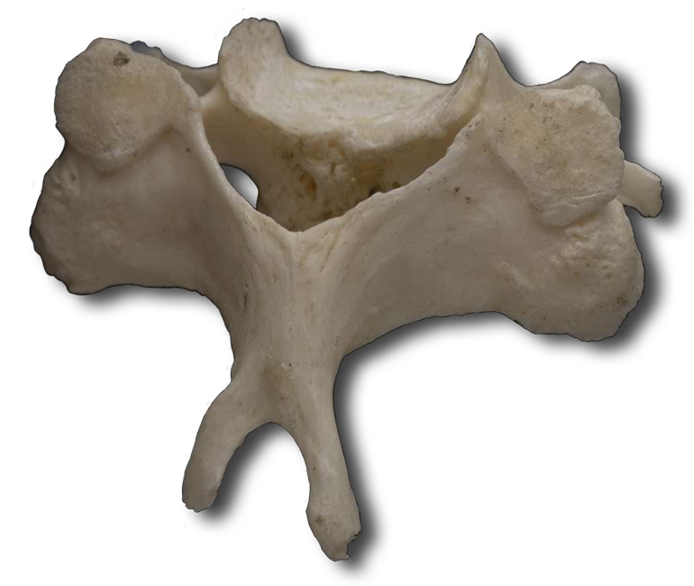
As you can see, thoracic vertebrae are considerably larger and stronger than cervical vertebrae. Rotate your specimen to observe the following:

Due to the amount of body weight supported by these vertebrae, they are the largest and strongest of the vertebral column. As always, rotate your specimen for the best observation angle.
From C2 on down, intervertebral discs separate each vertebra all the way down to the sacrum (which you will examine next). These discs help hold the vertebrae together while protecting the bone by preventing adjacent vertebrae from rubbing together and by absorbing vertical shock. The discs allow slight movements between adjacent vertebrae which translates into significant movement along the entire length of the column.
An intervertebral disc may be "herniated" if weakened or damaged in the performance of its shock absorbing function. This is sometimes referred to as a "slipped disc". This occurs when the annulus fibrosus is ruptured resulting in posterior protrusion of the nucleus pulposus. This is most common in the heavy weight bearing lumbar region. The consequence is pressure on the local spinal nerves which then radiates to other areas. Commonly, resulting pressure is exerted on the sciatic nerve resulting in pain radiating down the posterior thigh, through the calf, and even into the foot.
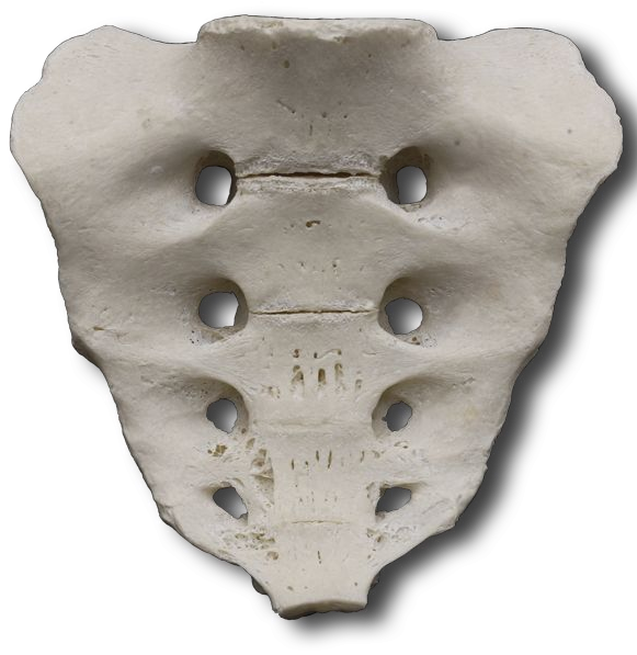
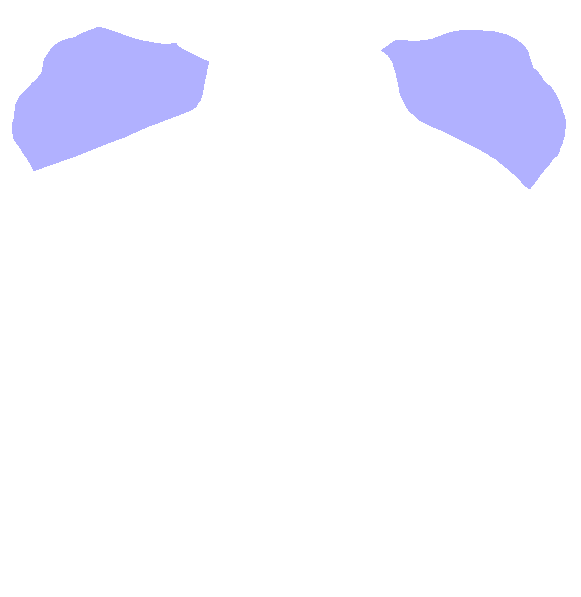
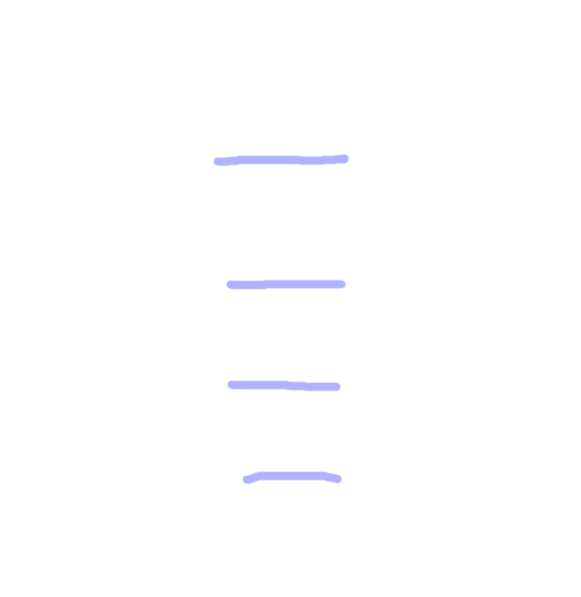
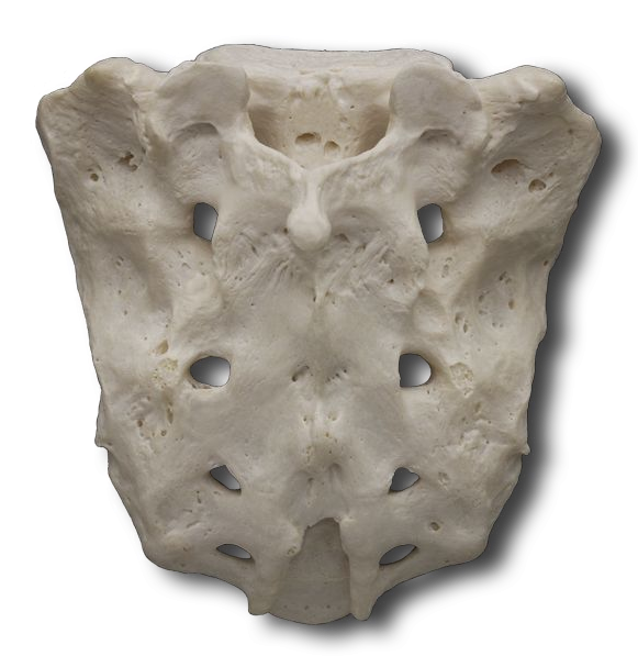
This triangularly shaped bone is formed by the fusion of five sacral vertebrae beginning between the ages of 16 and 18 years of age and usually completed by the age of 30. The sacrum is the support foundation for the pelvic girdle. To facilitate childbirth, the female sacrum is shorter, wider, and more curved than the male sacrum. Locate the following on your specimen and label them on your worksheet:
Known commonly as our tailbone, the coccyx is formed between the ages of 20 and 30 from the fusion of usually four coccygeal vertebrae. The coccyx is connected to the sacral cornu by ligaments and also is the connection point for other posterior muscles.
Let's take one last look at this entire structure. Identify each of the bones we've examined. Rotate your specimen to get a good perspective.