Expose and identify the spinal cord and its associated meninges.
Important Relationship
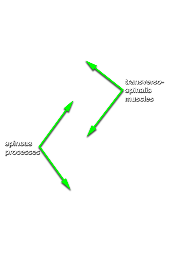
The erector spinae muscles are positioned superficial to the transversospinalis muscles.
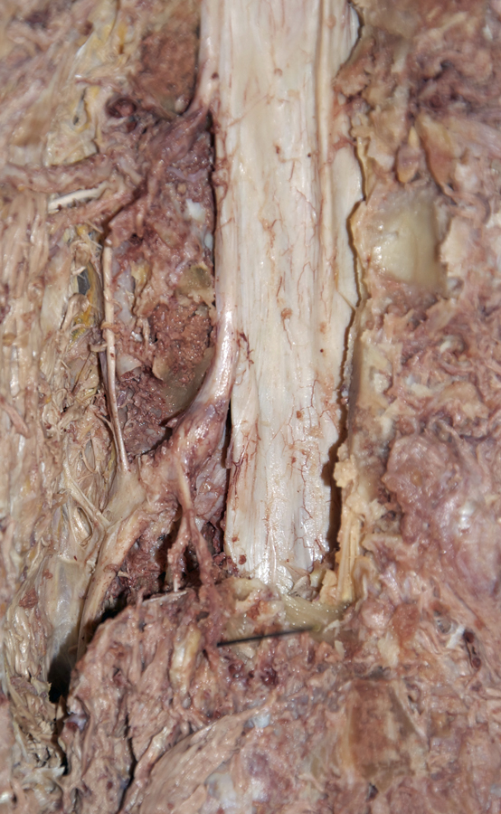
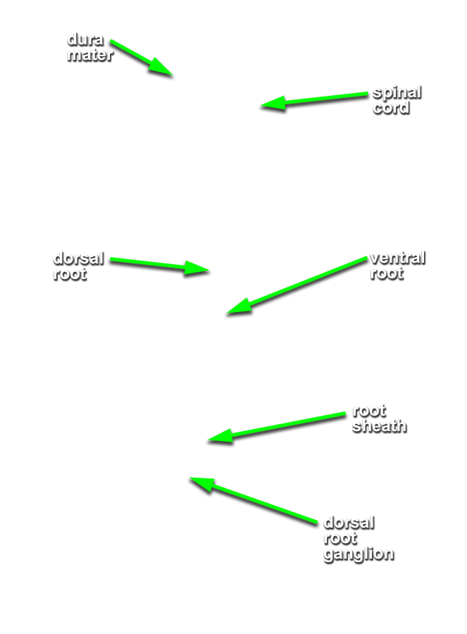
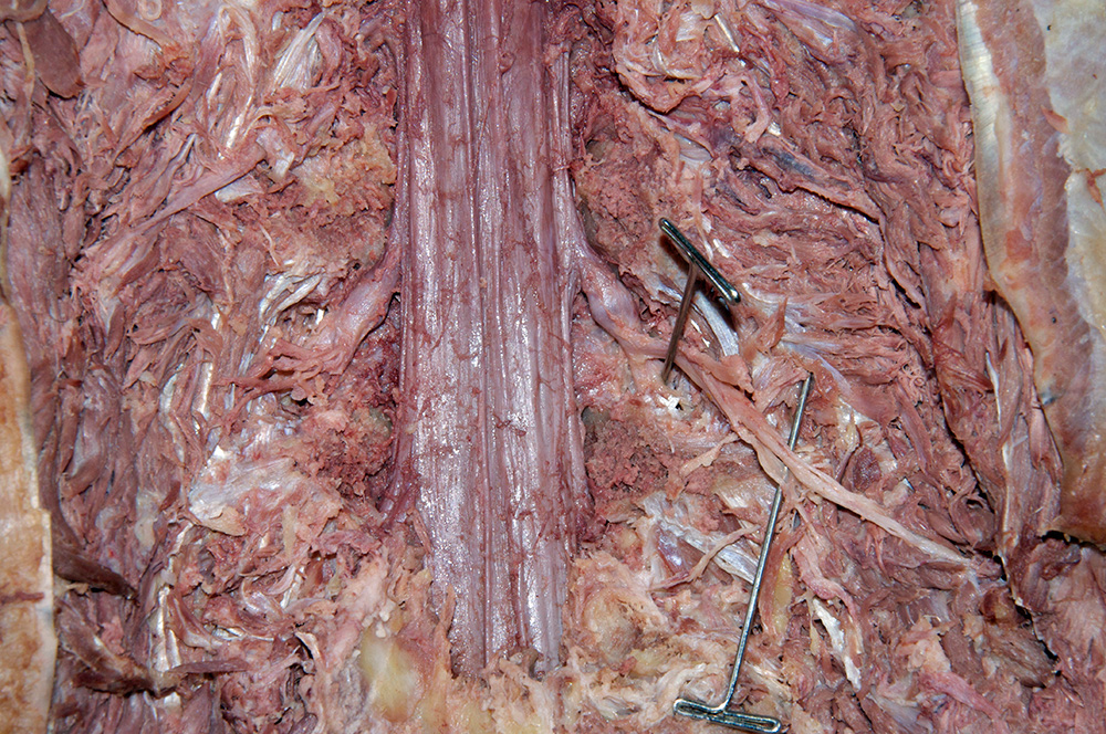
- Reflect the erector spinae muscle in the superior direction. The transversospinalis muscles and vertebral neural arches have been removed. Identify the dura mater surrounding the spinal cord. (G 1.37;N 165;Gl 4.7)
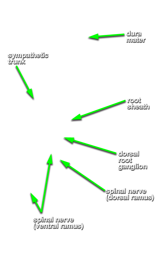
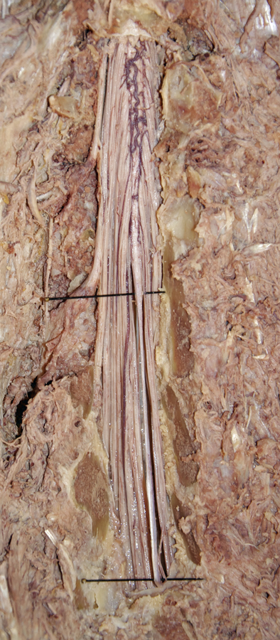
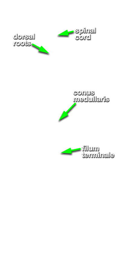
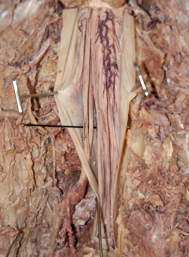
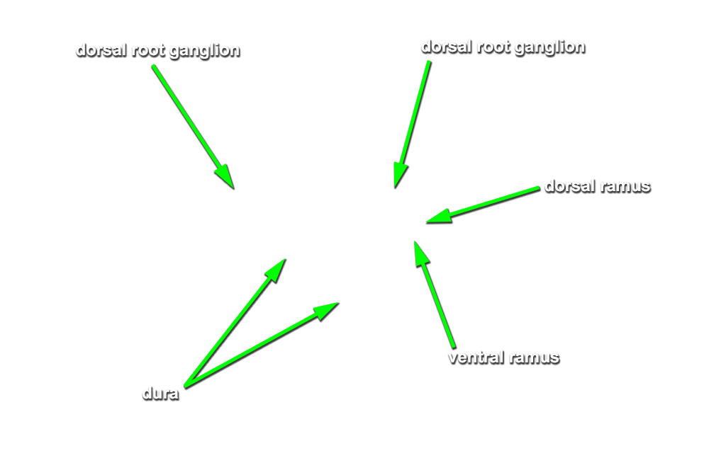
- Reflect the dura mater laterally and identify the arachnoid mater, spinal cord, dorsal and ventral roots (observe the dorsal and ventral roots in the cross section. Move through the cross sections 1 mm at a time and trace the dorsal and ventral roots to the dorsal root ganglion), conus medullaris, (follow the cross sections inferiorly in 1 mm steps to the tip of the conus medularis) cauda equina (no stucture is identified in the 3D Dissection Area as the cauda equina BUT the impression that the lumbar and sacral spinal nerve roots resemble a horse's tail is apparent in both the dissection and cross section areas) and filum terminale. (The filum terminale is not labeled as such. The spinal cord highlighted in slice 1651 may be the filum terminale. It will continue down to the sacrum. Observe the cross sections both superiorly into the conus medularis and inferiorly from slice 1651 to follow the non-labeled filum terminale to the sacrum.) (G 1.38;N 165;Gl 4.9)
- Identify the dorsal root ganglion, if it has been exposed