Identify and clean the structures of the submandibular and submental triangles.
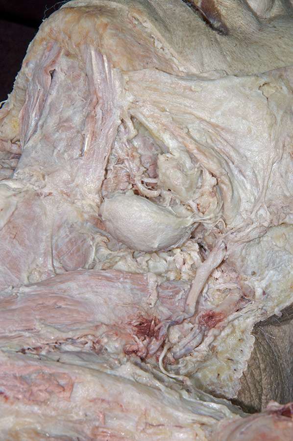
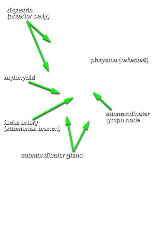
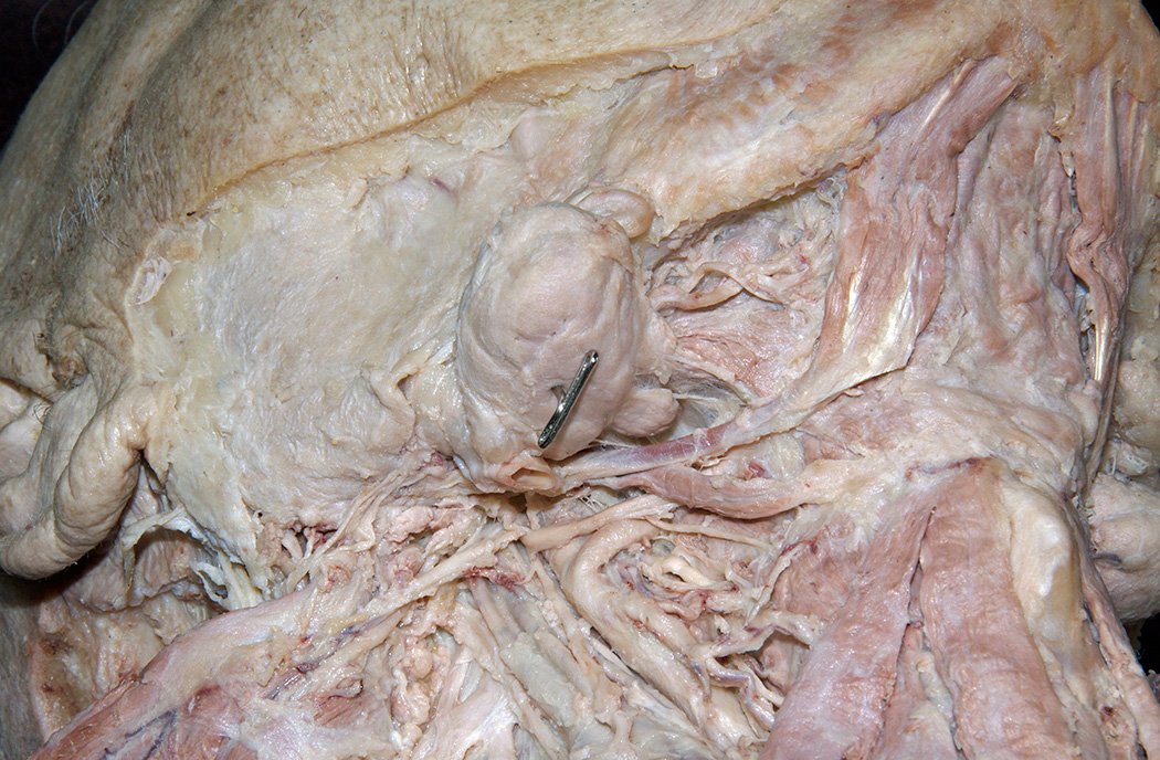
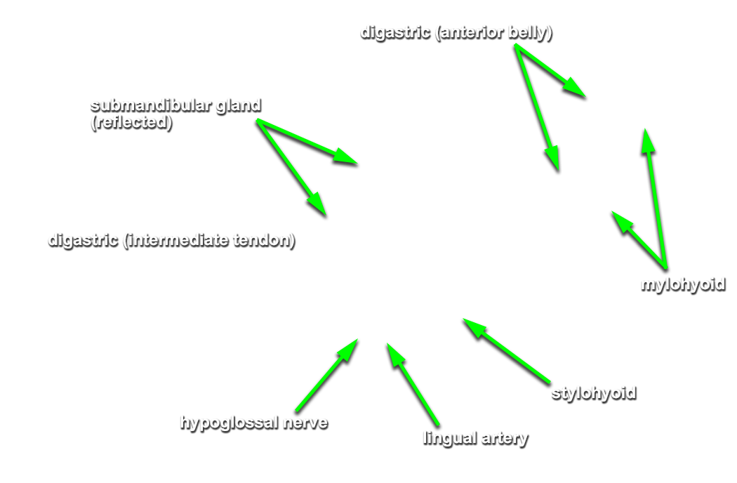
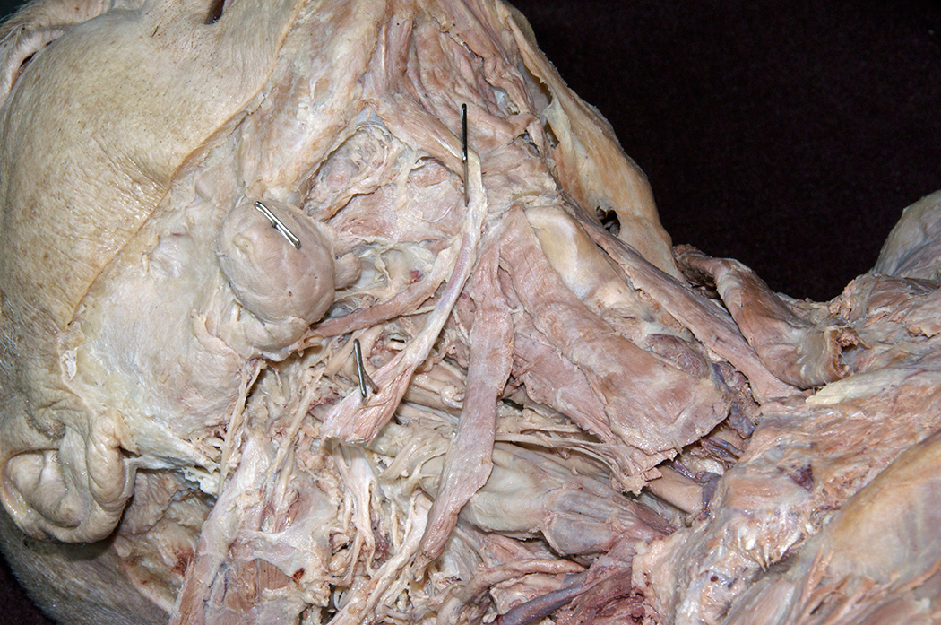
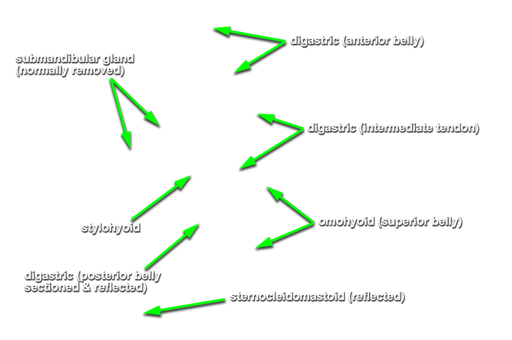
- (ON BOTH SIDES) Identify the submandibular salivary gland. (G 8.13;N 46;Gl 44.25A)
- (ON THE RIGHT SIDE ONLY) Trace (blunt dissect) the facial artery and vein through the gland. Remove the gland as you isolate the facial artery and vein (you will identify the origin of the facial artery in a subsequent step).
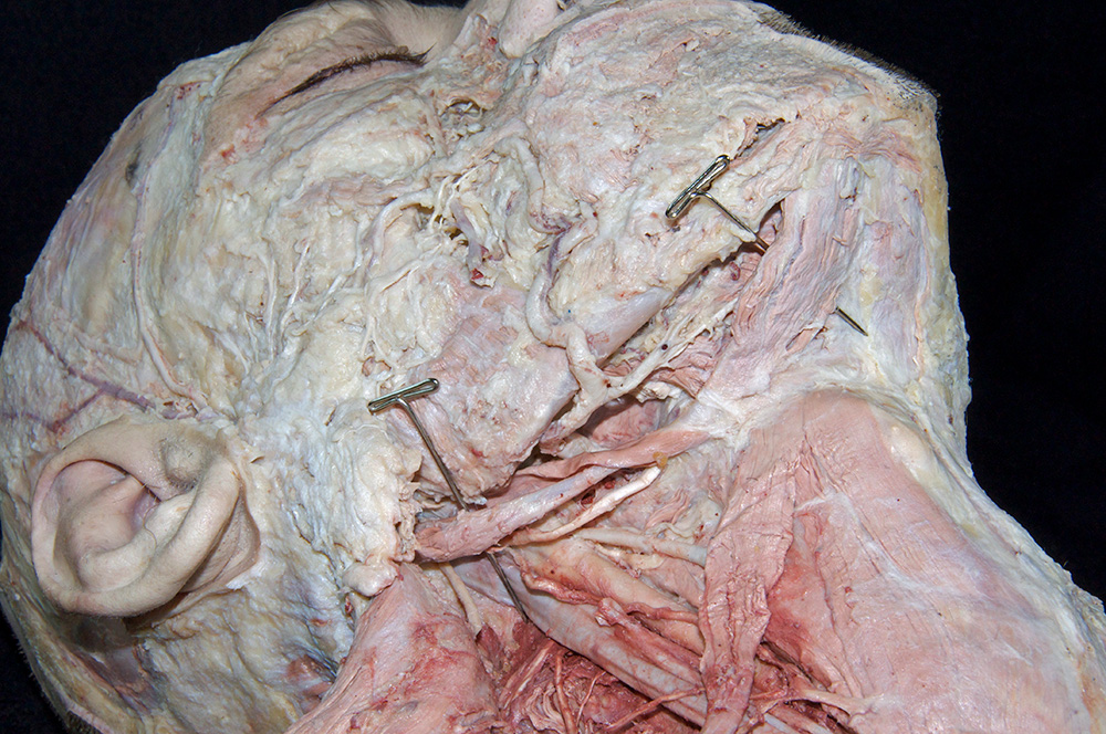
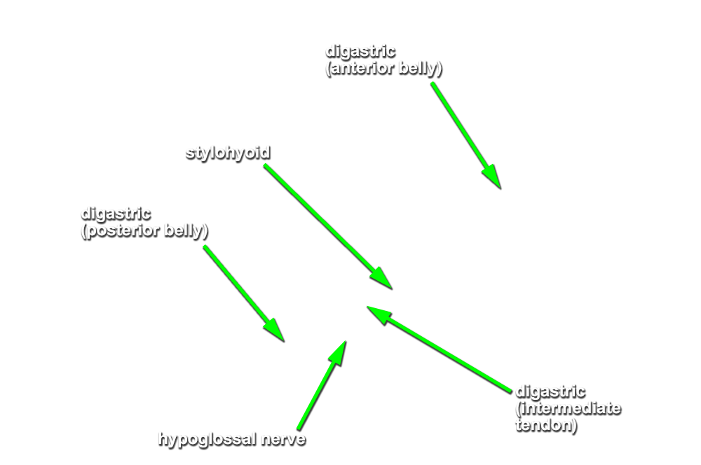
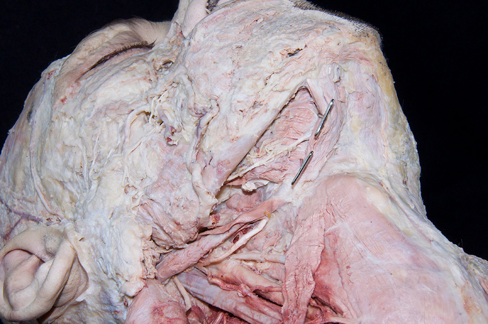
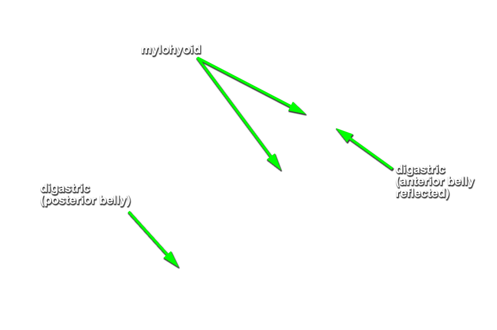
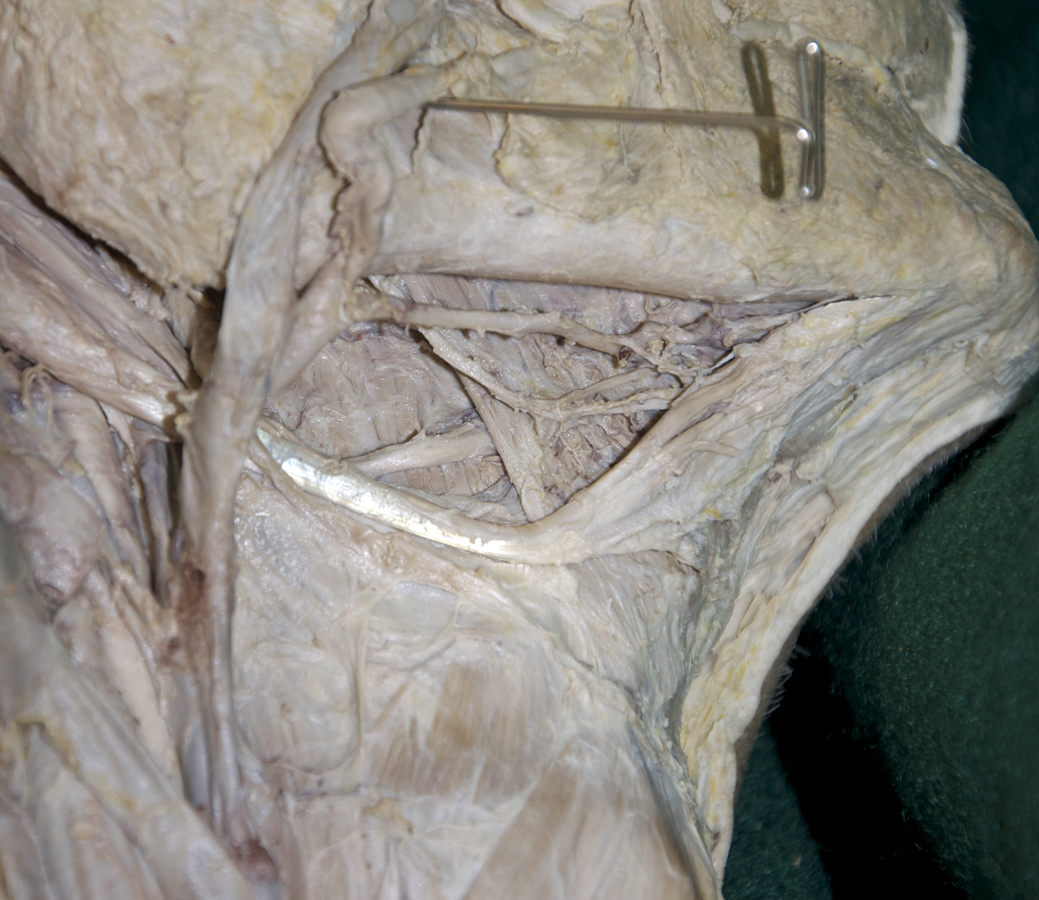
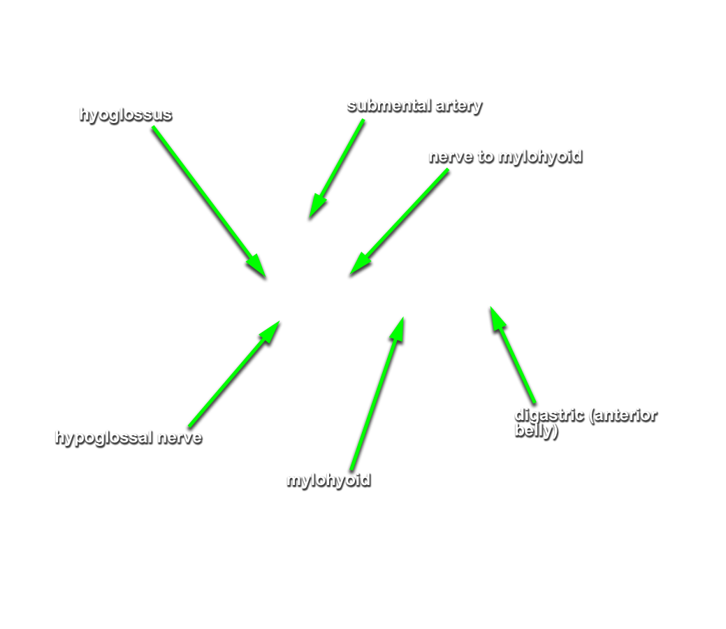
- (ON BOTH SIDES) Identify and clean the anterior and posterior bellies of the digastric muscle, the intermediate tendon of the digastric muscle, and the stylohyoid and mylohyoid muscles. (G 8.14;N 29;Gl 44.13) Attempt to identify the nerve to the mylohyoid.
- (ON THE RIGHT SIDE ONLY) Trace and clean the posterior belly of the digastric to its proximal attachment. Remove any remaining parotid gland but do not remove the stylohyoid muscle or any of the nerves or vessels. Cut the posterior belly of the digastric at its proximal attachment. Leave the muscle belly attached to its intermediate tendon.
Important Relationships
- The submandibular gland is positioned posterior and inferior to the mylohyoid muscle.
- The digastric muscle (anterior belly) is positioned superficial (inferior-lateral) to the mylohyoid muscle.