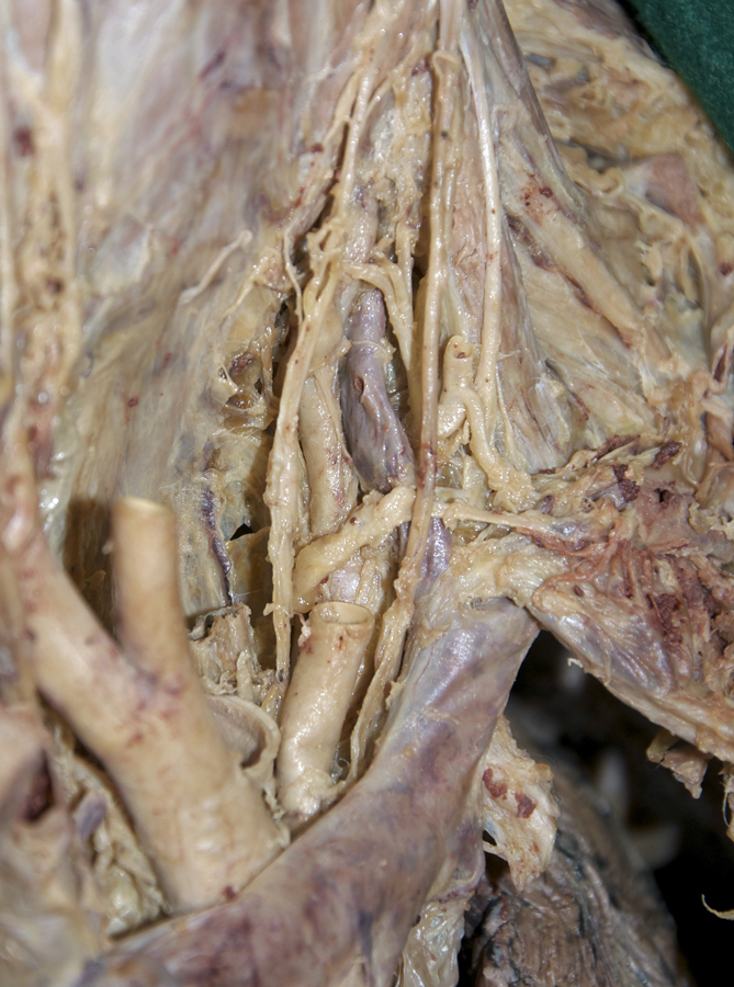
Identify and clean the deep structures of the anterior neck.
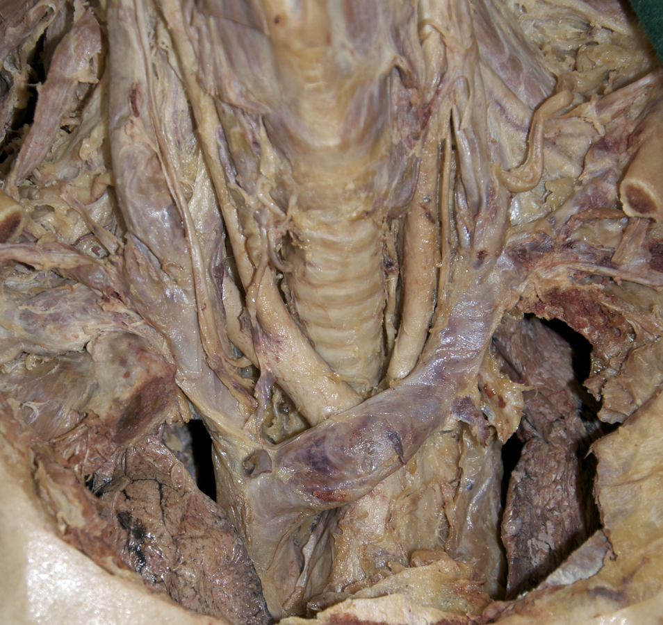
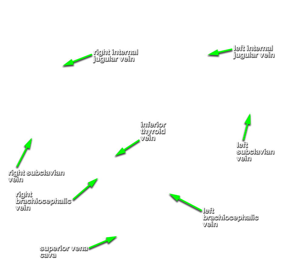
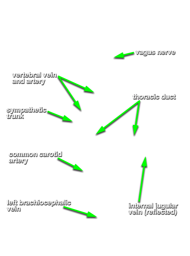
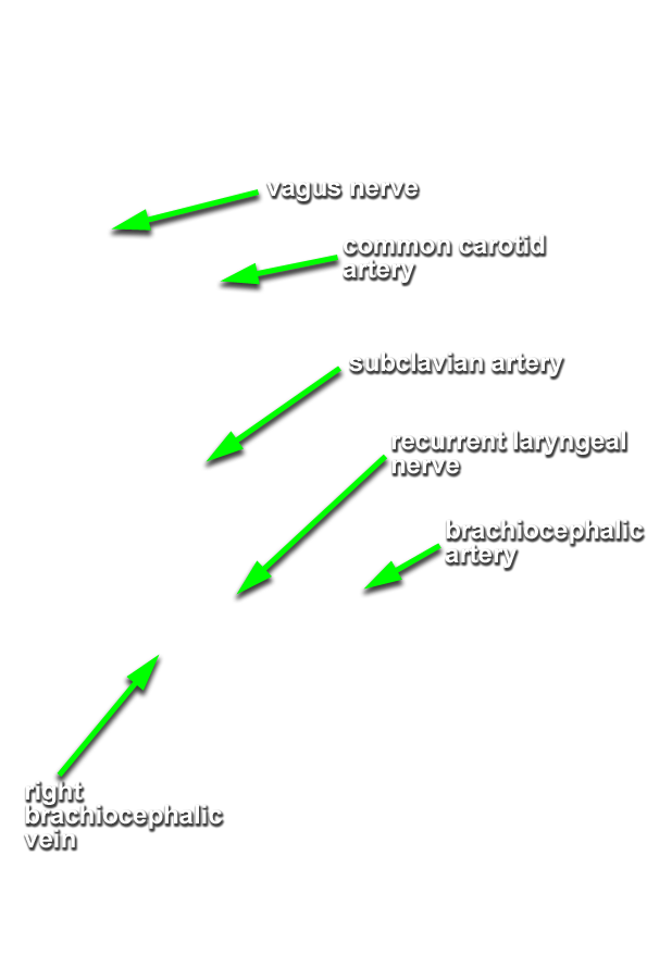
- (ON BOTH SIDES) Identify the brachiocephalic veins and the brachiocephalic artery. (G 8.22B;N 203;Gl 9.6A) Transect the common carotid artery (same level as the internal jugular vein) and reflect (do not remove) the artery in the superior and inferior directions. Make certain that you do not cut the vagus nerve.
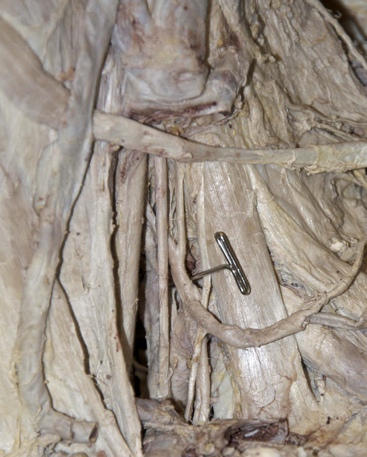
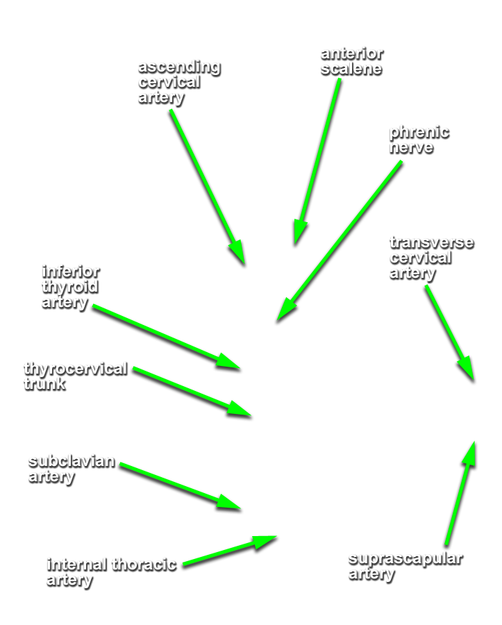
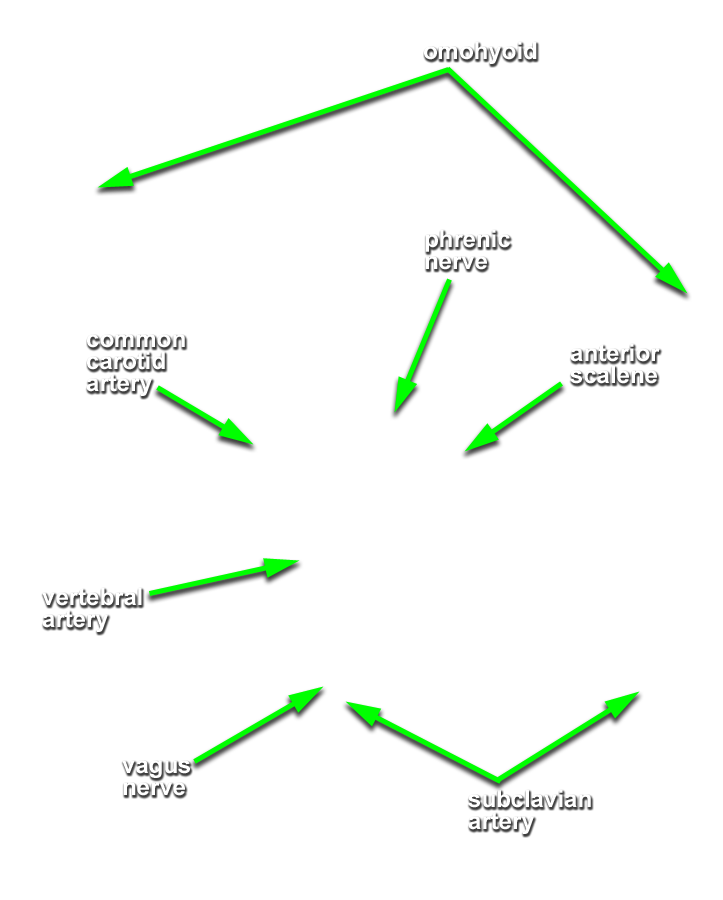
- (ON BOTH SIDES) Return to the transverse cervical and suprascapular arteries. Trace these arteries in the proximal direction back towards their origin. Identify the thyrocervical trunk. (G 8.22A;N 33;Gl 45.31C) Identify and clean the inferior thyroid and ascending cervical arteries. Trace the inferior thyroid artery to the thyroid gland.
- The left brachiocephalic vein passes anterior to both the left common carotid and brachiocephalic arteries.
- The left phrenic nerve passes posterior to the left brachiocephalic vein.
- The inferior thyroid artery passes deep (posterior-medial) to the common carotid artery.
- The ascending cervical artery is positioned directly anterior to the anterior scalene muscle.
- (ON BOTH SIDES) Trace the thyrocervical trunk to the subclavian artery. Identify the internal thoracic and vertebral arteries. (G 8.22B;N 33;Gl 45.31D) On the left side of the cadaver, identify the thoracic duct where it drains into the brachiocephalic vein. The thoracic duct passes posterior to the arch of the aorta and origin of the common carotid artery before passing anterior to the vertebral artery and thyrocervical trunk. It is often filled with blood where it enters the brachiocephalic vein and appears like a small venous tributary.
- The thoracic duct passes anterior to the left subclavian artery and phrenic nerve, and posterior to the common carotid artery, vagus nerve and internal jugular vein.
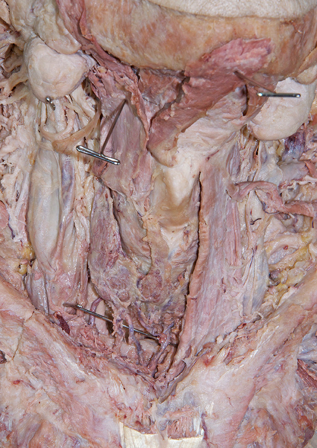
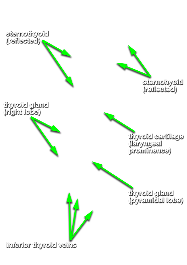
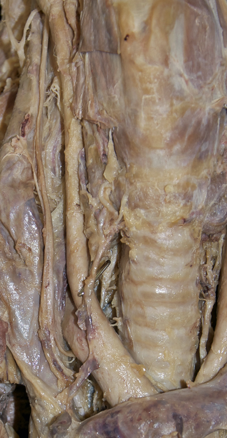
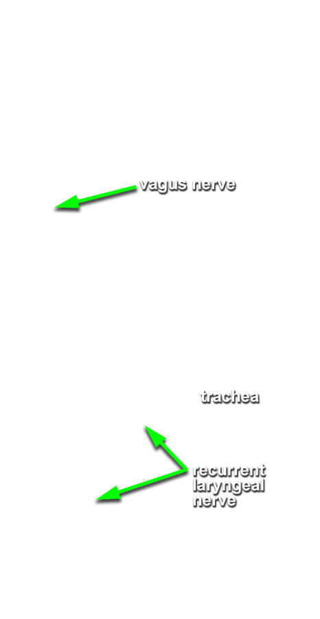
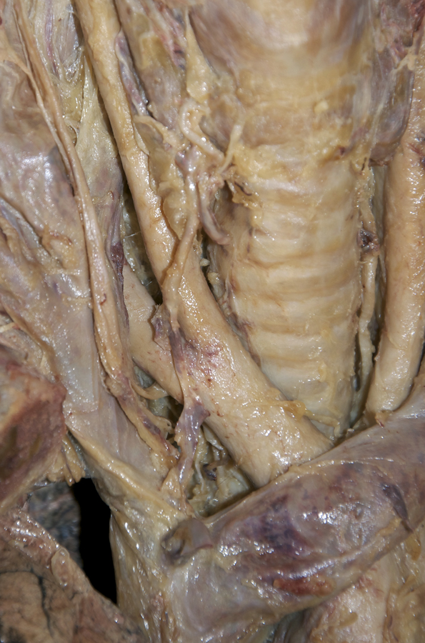
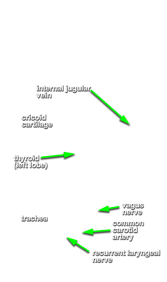
- (ON THE RIGHT SIDE ONLY) Return to the thyroid gland. Identify the inferior thyroid vein(s). (G 8.21;N 76;Gl 45.31C) Blunt dissect around and then remove the right lobe of the thyroid gland. Attempt to identify the parathyroid glands. Blunt dissect in the tracheoesophageal groove and identify the recurrent laryngeal nerve. Trace the recurrent layngeal nerve to its origin from the vagus nerve. (G 8.22A;N 77;Gl 45.26) Attempt to identify the left recurrent laryngeal nerve without removing the left lobe of the thyroid gland.
- The trachea is positioned directly anterior to the esophagus.
- The recurrent laryngeal nerve is positioned lateral to the trachea.
- The right recurrent laryngeal nerve passes inferior and posterior to the right subclavian artery.
- The vagus nerves pass directly anterior to the subclavian arteries.
Important Relationships
Important Relationship
Important Relationships