If necessary, remove the remaining anterior (proximal) portion of the lateral pterygoid muscle to expose the medial pterygoid muscle, and nerves, arteries and veins of the infratemporal fossa.
- (ON THE RIGHT SIDE ONLY) If necessary, carefully shred the lateral pterygoid muscle. Use forceps and scissors to remove the shredded muscle.
- (ON THE RIGHT SIDE ONLY) Identify and clean the medial pterygoid muscle. (G 7.50B;N 49;Gl 40.26)
-
(ON THE RIGHT SIDE ONLY) Identify and clean the
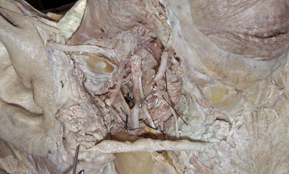
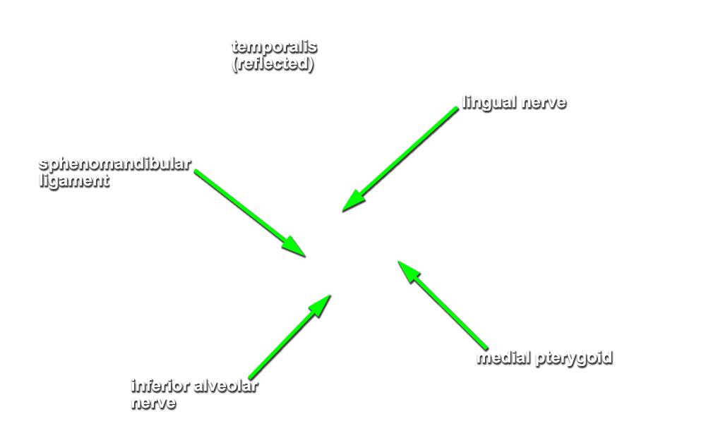
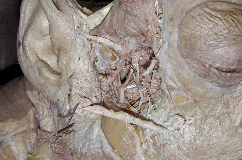
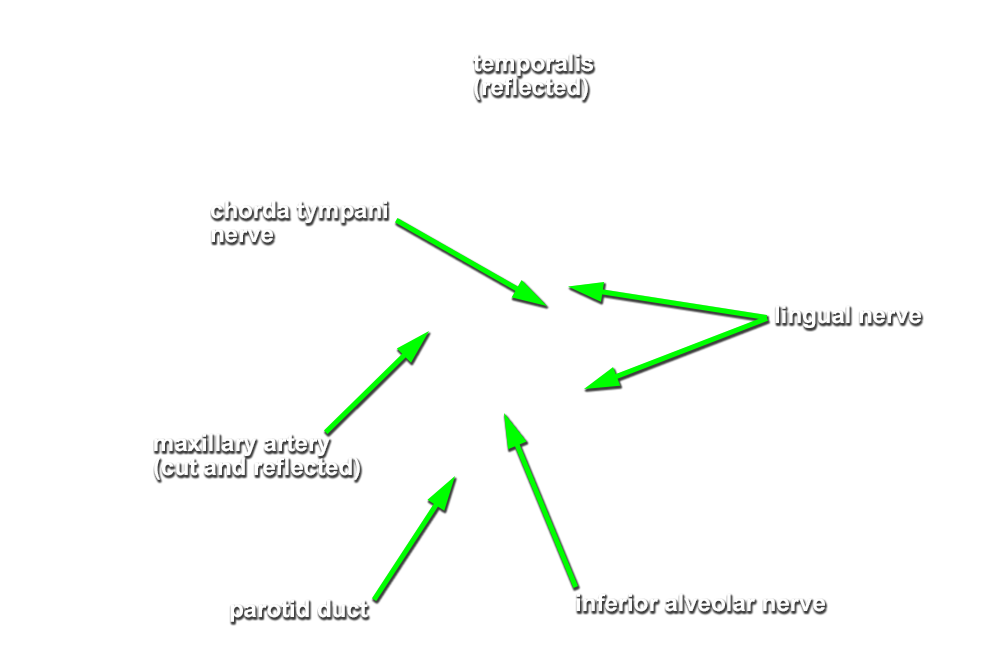
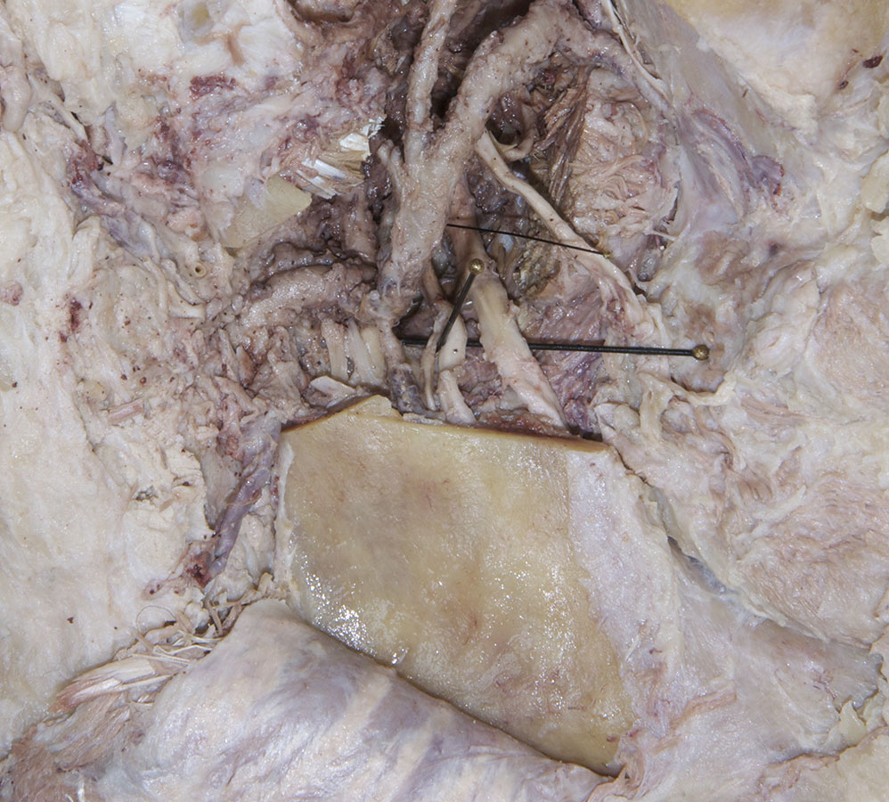
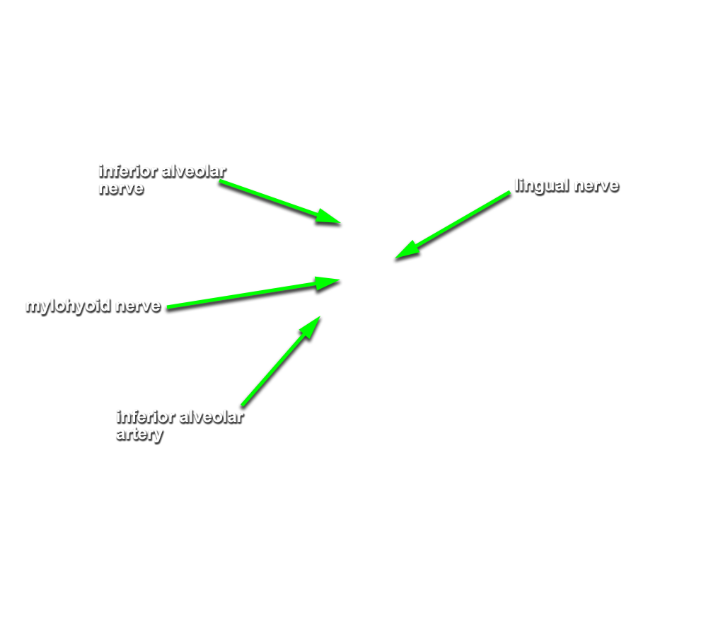
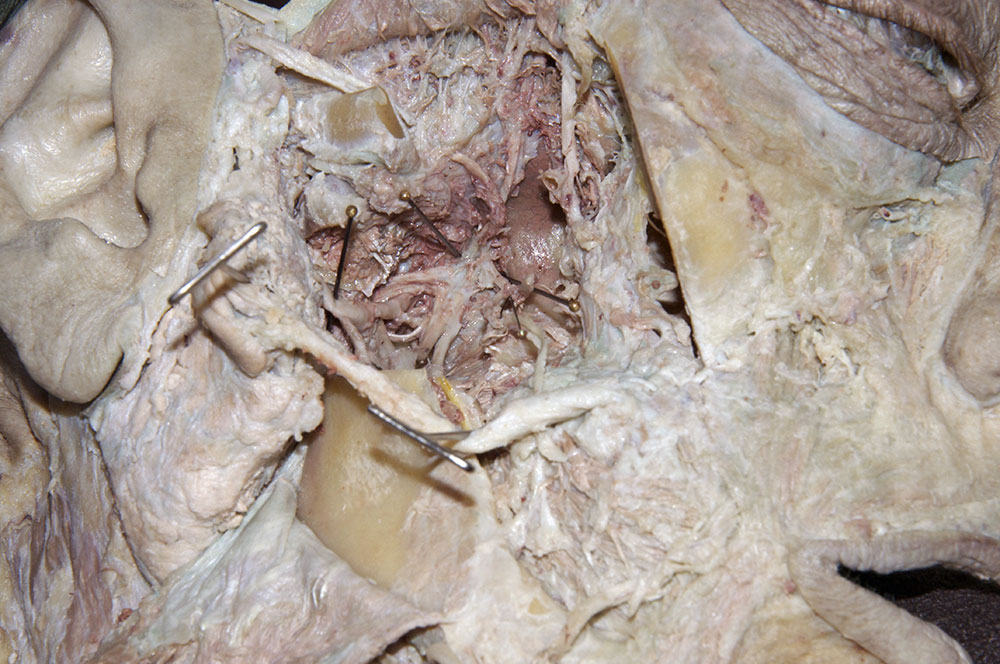
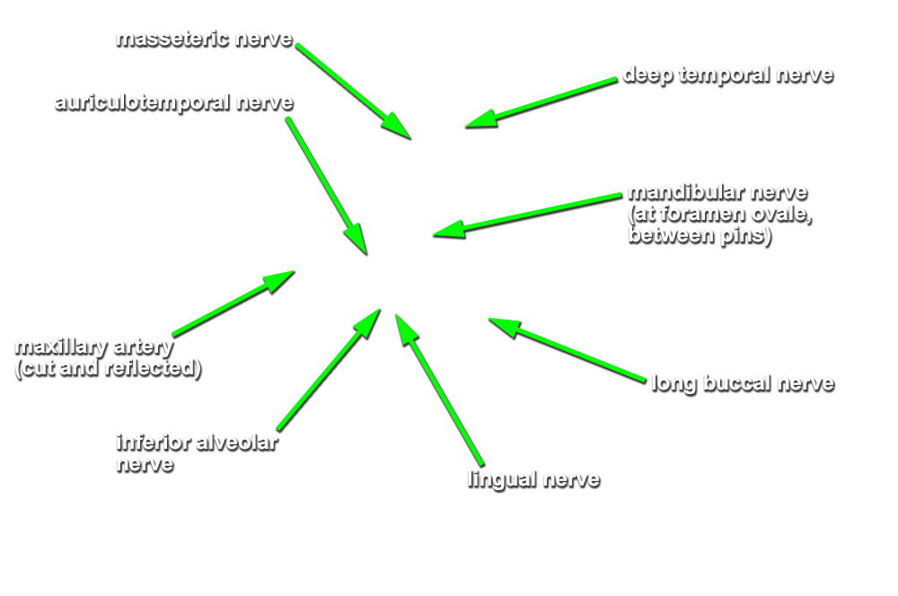
inferior alveolar and
lingual nerves and the
sphenomandibular ligament. (G 7.50B;N 51;Gl 44.5) Attempt to identify the
chorda tympani nerve and the
nerve to the mylohyoid muscle.
Important Relationships
- The lingual nerve passes medial to the mandible and lateral to the medial pterygoid muscle and is positioned anterior to the inferior alveolar nerve.
-
(ON THE RIGHT SIDE ONLY) Identify the
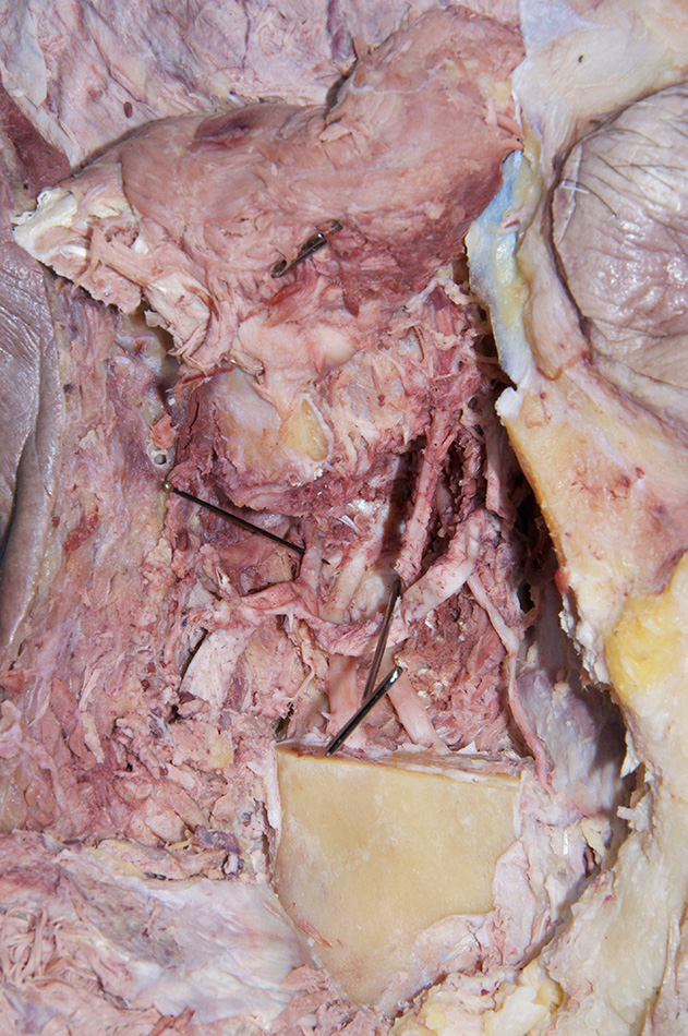
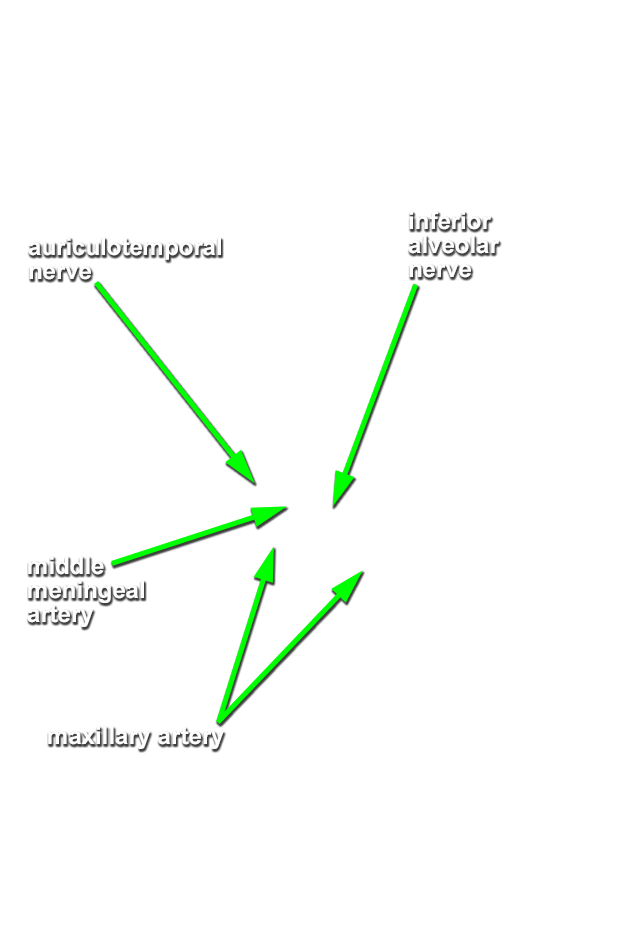
inferior alveolar artery. (G 7.51;N 51;Gl 40.26) This artery should be with the inferior alveolar nerve. Clean and trace the inferior alveolar artery in the superior direction until you reach the
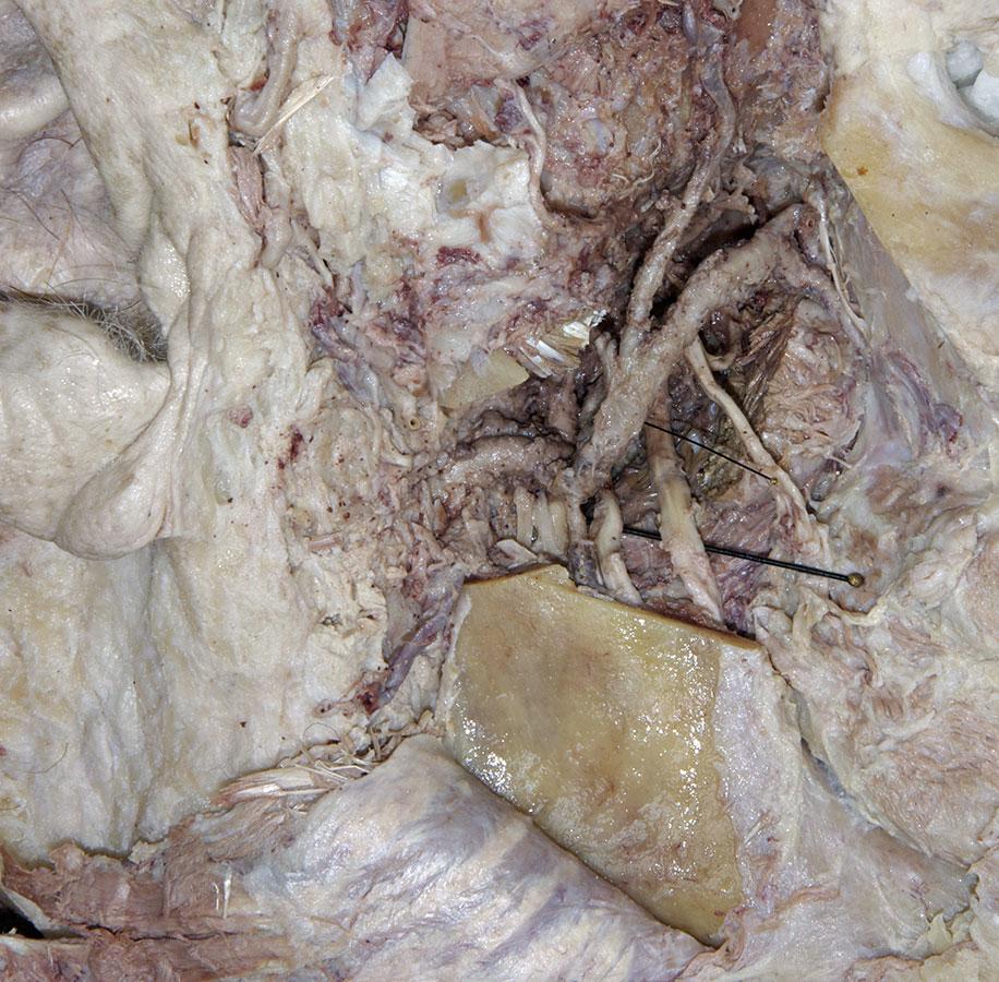
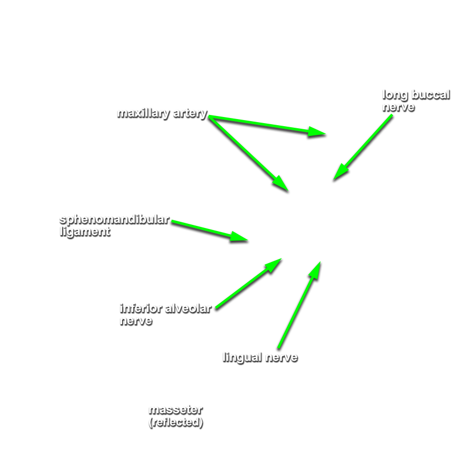
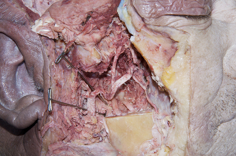
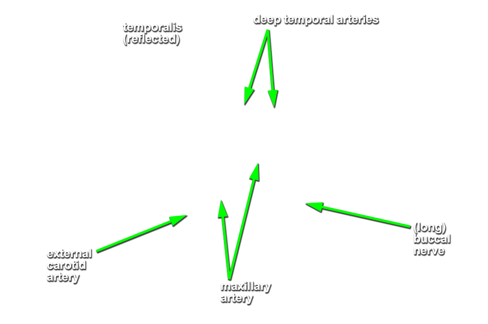
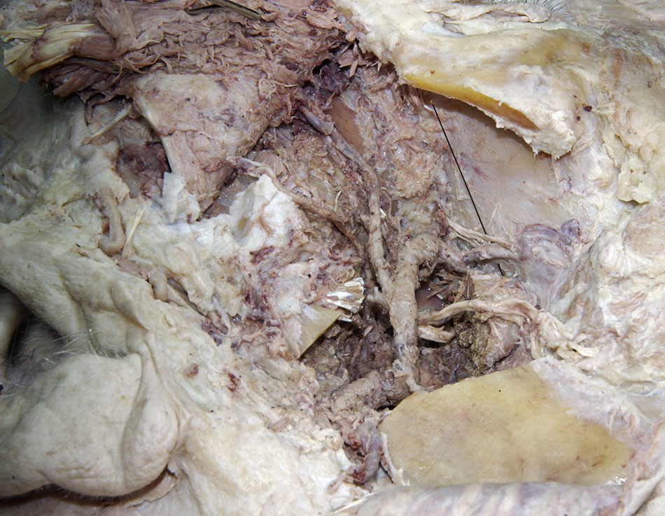
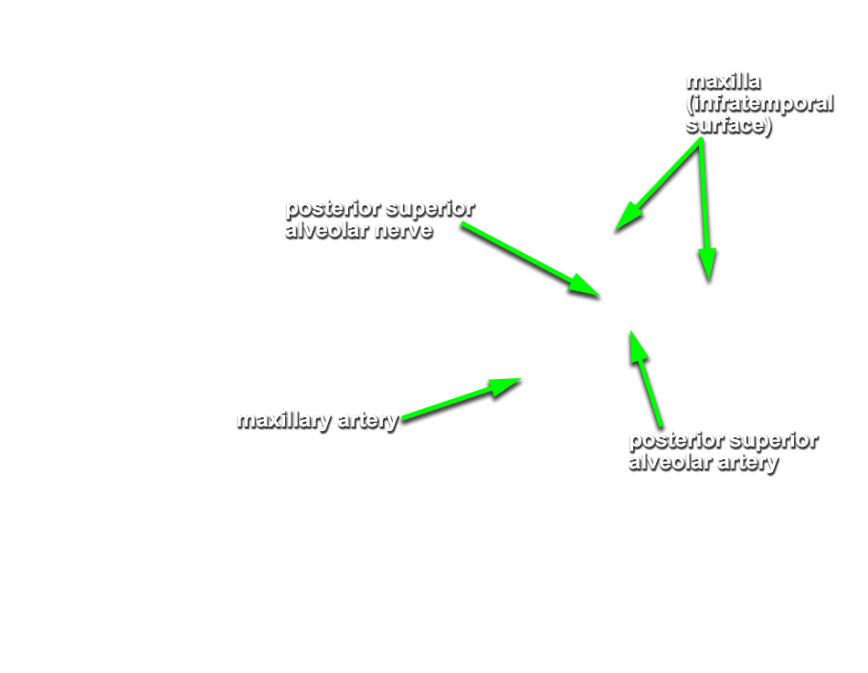
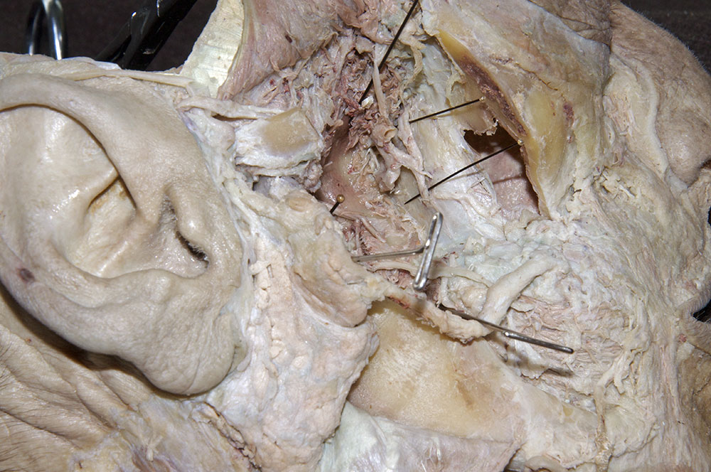
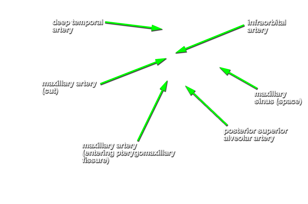
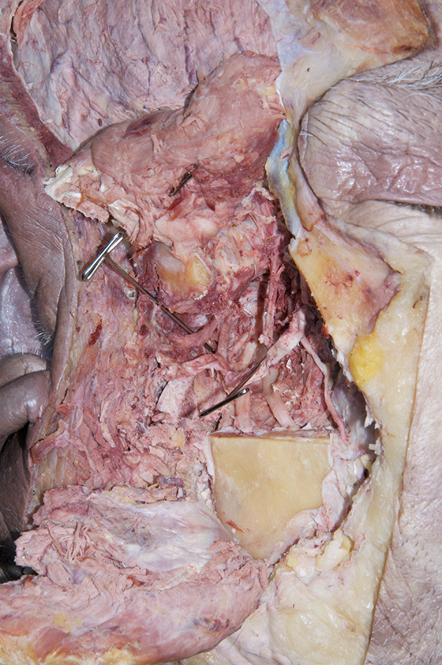
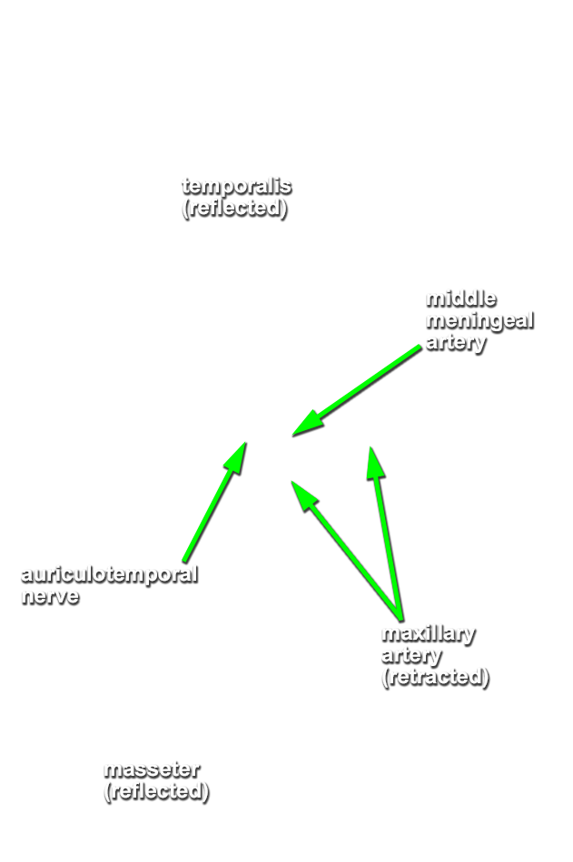
maxillary artery. Clean and trace the maxillary artery medially. As you clean the maxillary artery, identify the
middle meningeal, deep temporal, and posterior superior alveolar arteries. Attempt to trace the maxillary artery to the pterygomaxillary fissure. Identify the
auriculotemporal nerve splitting around the middle meningeal artery. You may need to remove the condylar process of the mandible and remaining distal lateral pterygoid muscle to expose the middle meningeal artery and auriculotemporal nerve. Identify the
(long) buccal nerve. Attempt to trace the long buccal, auriculotemporal, lingual, and inferior alveolar nerves back to foramen ovale.
Important Relationships
- The medial pterygoid muscle is positioned medial (deep) to the mandible (ramus).
- The maxillary artery passes medial to the mandible (neck) and lateral to the sphenomandiblar ligament. It typically passes lateral to the lateral pterygoid muscle.
-
(ON THE RIGHT SIDE ONLY) Identify the
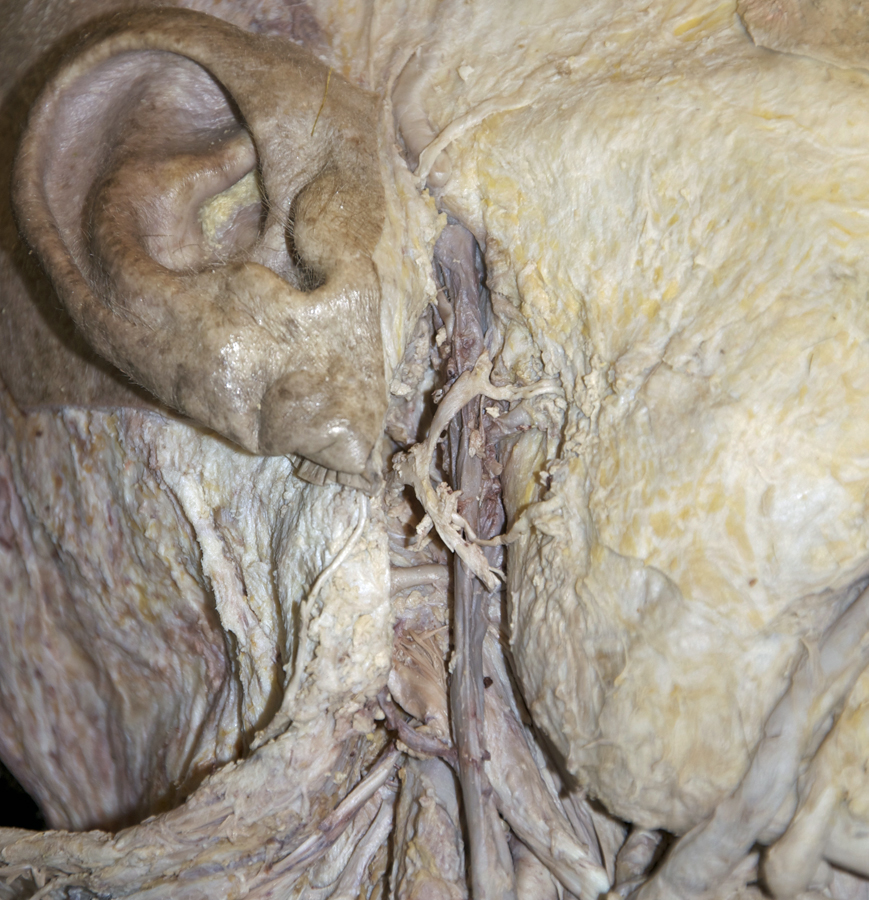
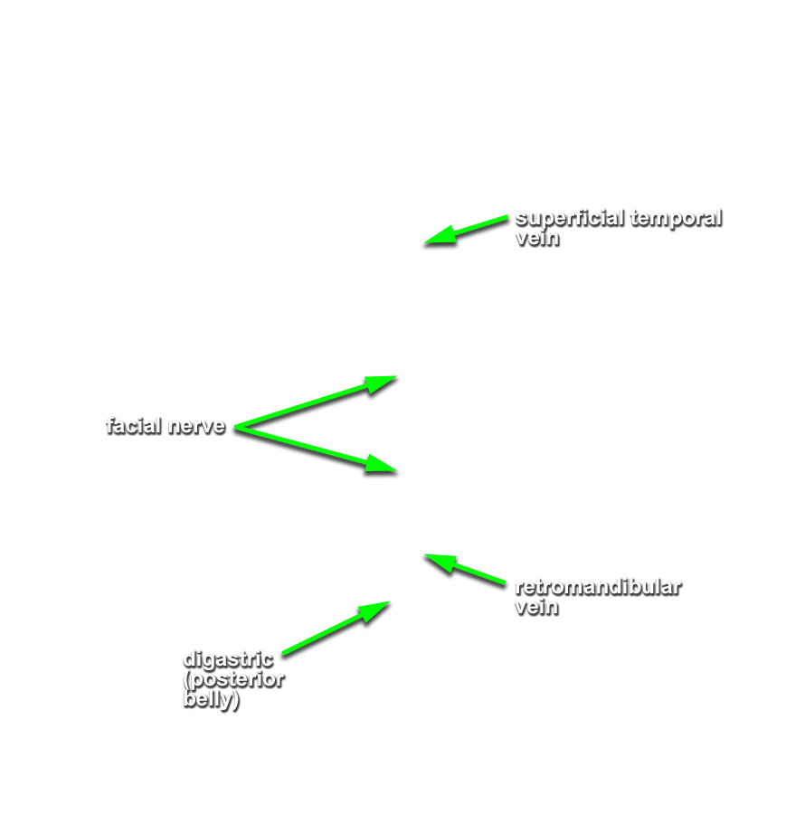
retromandibular vein. (G 7.47C;N 73;Gl 40.10) Trace the retromandibular vein in the inferior direction until it drains into the
common facial vein (or external jugular vein). Remove any remaining parotid gland and be careful to leave the stump of the facial nerve. Attempt to leave the veins (retromandibular, external jugular, facial and common facial) intact.
Important Relationship
- The retromandibular vein is positioned posterior-medial to the mandible (ramus).