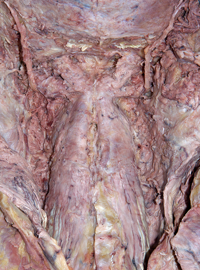
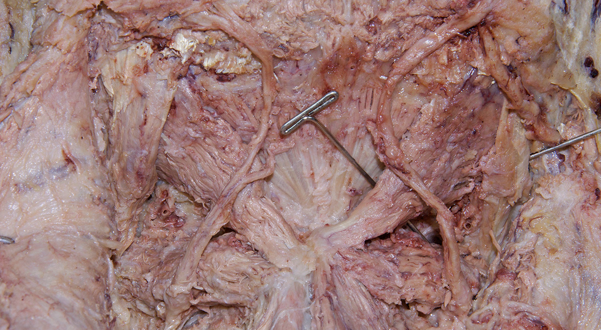
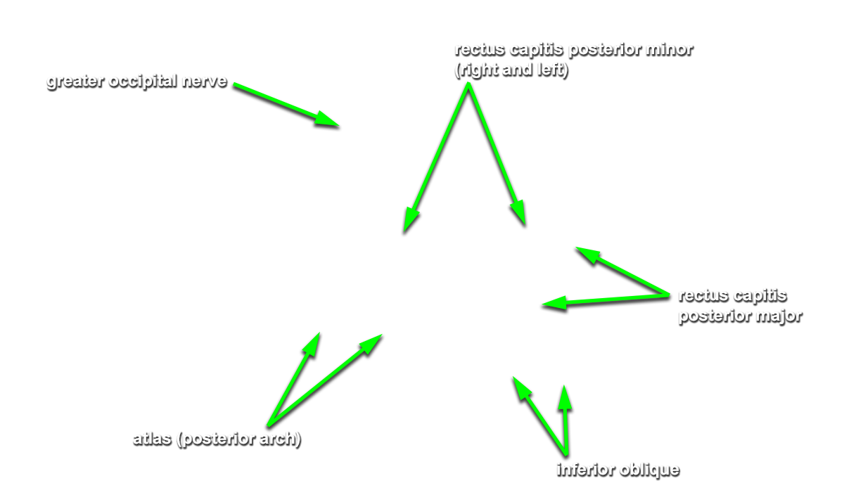
Identify and clean the structures of the suboccipital triangle. (G 1.34A;N 175;Gl 3.4)
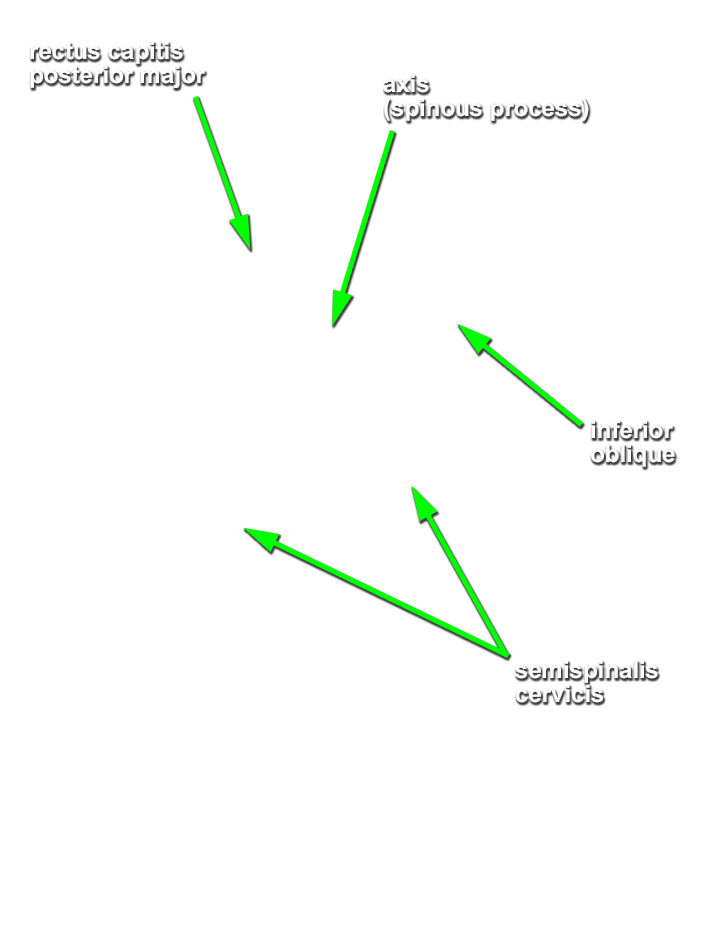
- Start by identifying (palpation) the bifid spinous process of the axis.
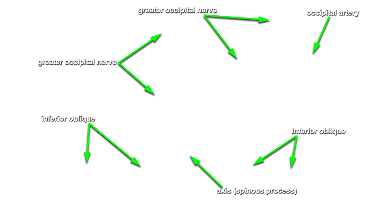
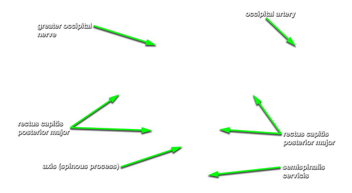
(ON BOTH SIDES) Identify the semispinalis cervicis muscle attaching to the spinous process of the axis. Use the spinous process of the axis and the greater occipital nerve to identify the inferior oblique muscle. The greater occipital nerve passes posterior and then inferior to this muscle. - (ON BOTH SIDES) Identify the rectus capitis posterior major muscle. This muscle also attaches to the spinous process of the axis.
- (ON BOTH SIDES) Return to the inferior oblique muscle. Trace and clean the inferior oblique muscle towards the transverse process of the atlas. Identify and clean the superior oblique muscle. This muscles attaches to the transverse process of the atlas and the occipital bone. Attempt to identify the suboccipital nerve in the suboccipital triangle.
-
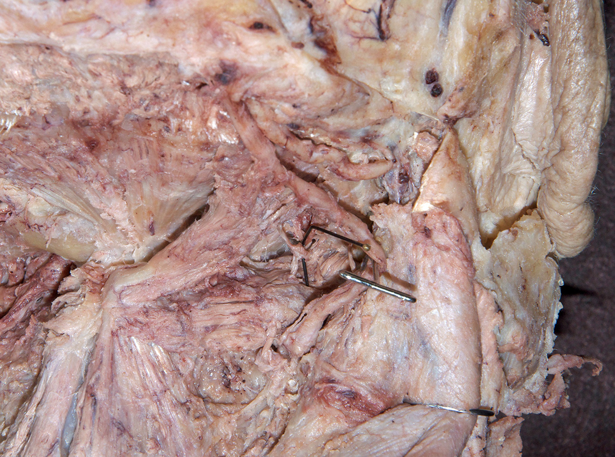
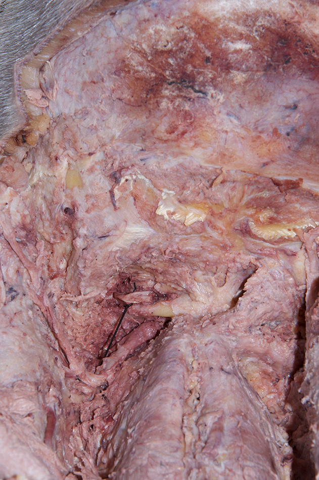
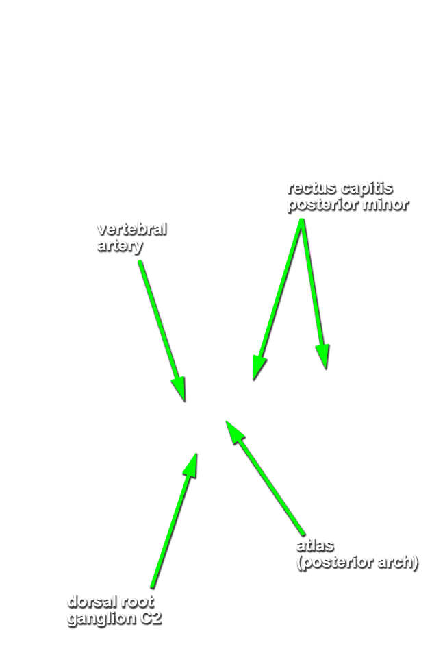
(ON ONE SIDE ONLY) Remove the rectus capitis posterior major and inferior oblique muscles. Do not damage the greater occipital nerve. Identify and clean the
rectus capitis posterior minor muscle and
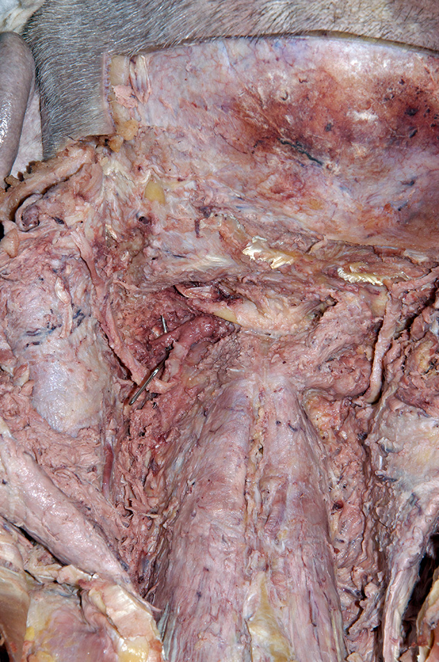
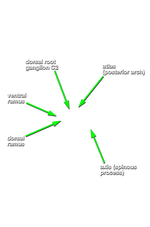
posterior arch of the atlas. Carefully blunt dissect (tear the posterior atlanto-occipital membrane) along the superior aspect of the posterior arch of the atlas to expose the
vertebral artery. (N 175; Gl 45.37) Trace (blunt dissect) the greater occipital nerve in the proximal direction to identify the
C2 dorsal root ganglion.
Important Relationship
- The greater occipital nerve passes inferior and posterior to the inferior oblique muscle.