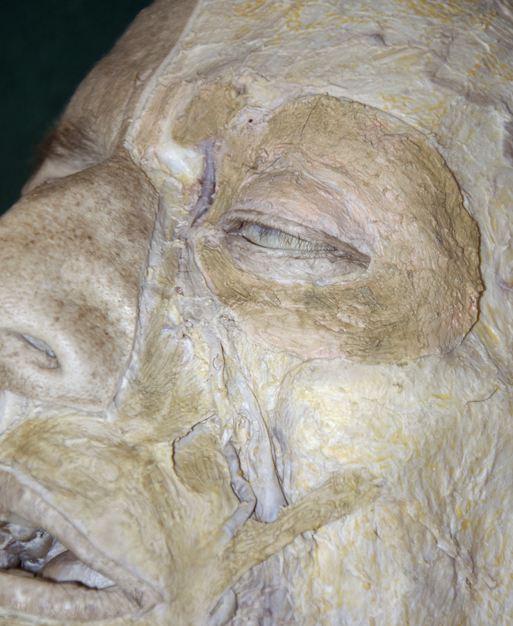
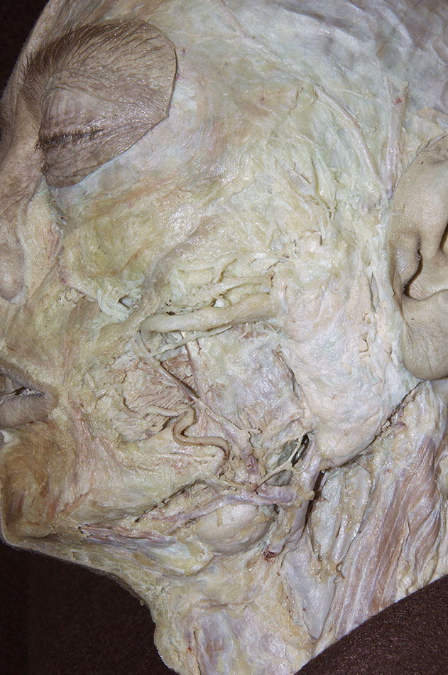
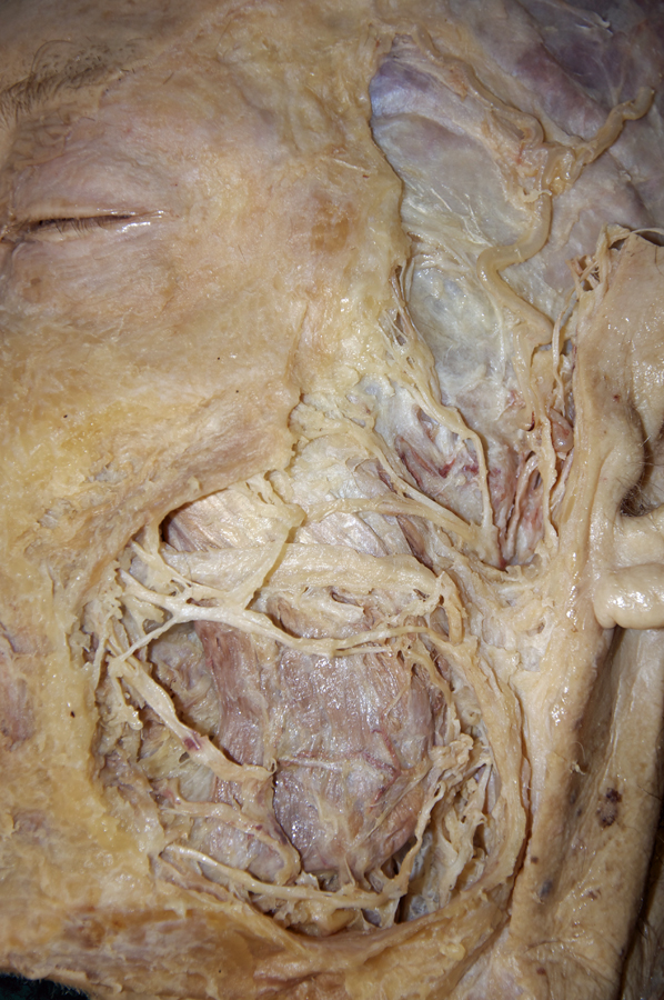
(ON THE LEFT SIDE ONLY) Identify the muscles of facial expression, the parotid gland and duct, and the branches of the facial nerve (refer to the interactive photo above for illustrations of the muscles of facial expression and the facial nerve).
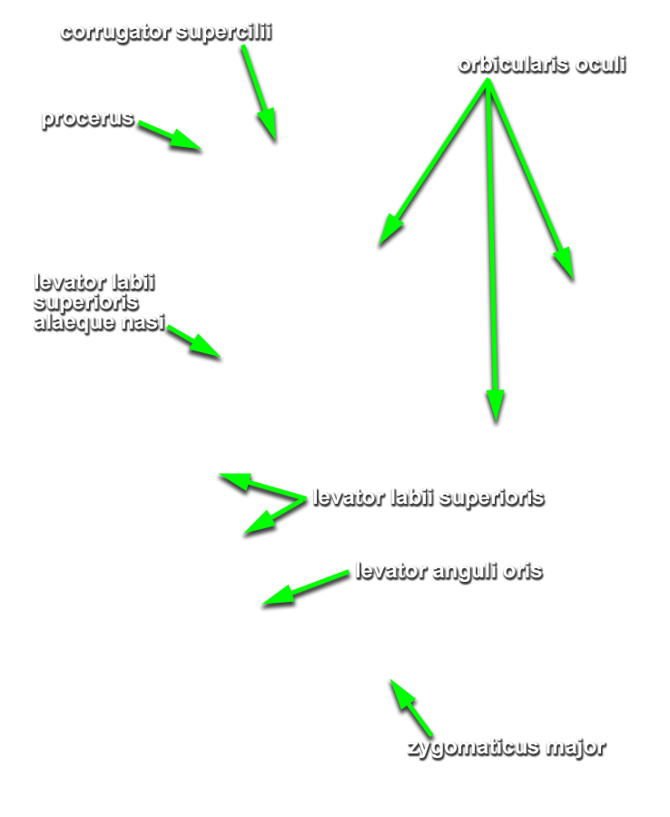
- Identify the frontalis and orbicularis oculi muscles. (G 7.12;N 25;G 38.1) Attempt to distinguish the orbital and palpebral parts of the orbicularis oculi. (N 25;Gl 38.1)
- Identify the
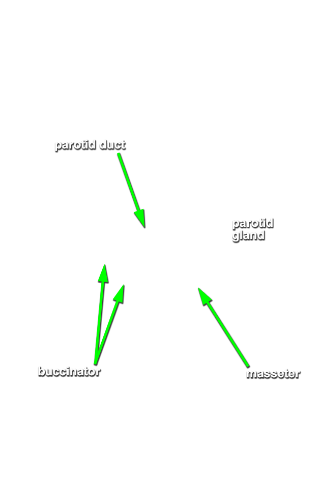
parotid gland and
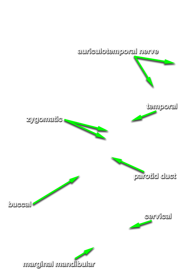
duct. (G 7.12;N 24;Gl 40.21) Note (in the interactive photograph) the orientation of the facial nerve branches adjacent to the parotid duct. Identify the
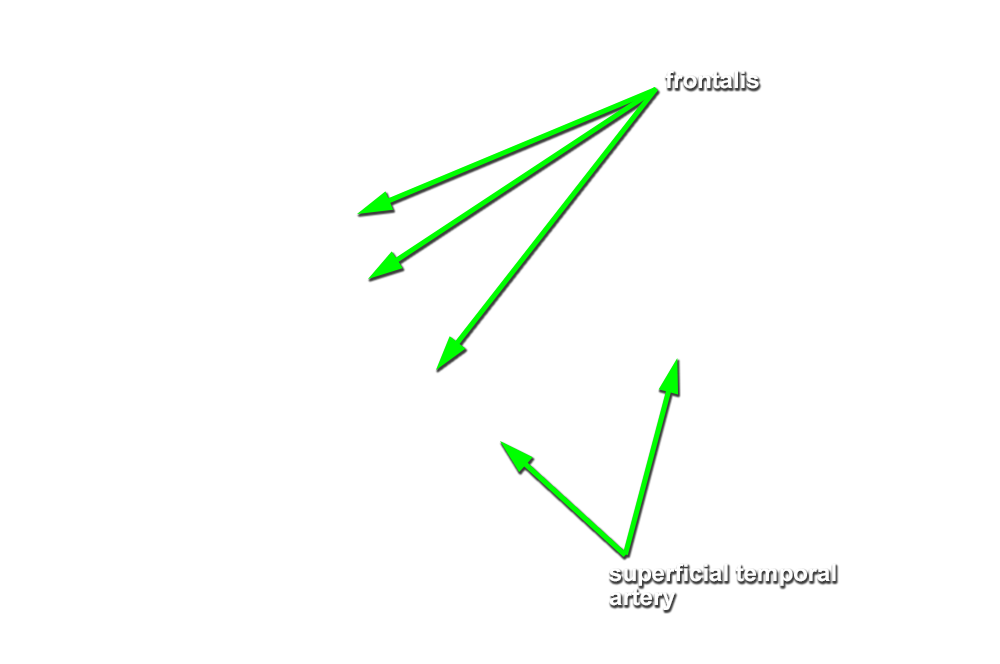
temporal,
zygomatic,
buccal,
mandibular and
cervical branches of the facial nerve. (G 7.13;N 24;Gl 40.21)
Important Relationships
- The parotid duct passes lateral (superficial) and anterior to the masseter muscle.
- The parotid gland is positioned posterior and lateral (superficial) to the masseter muscle.
- The branches of the facial nerve pass lateral (superficial) to the masseter muscle.
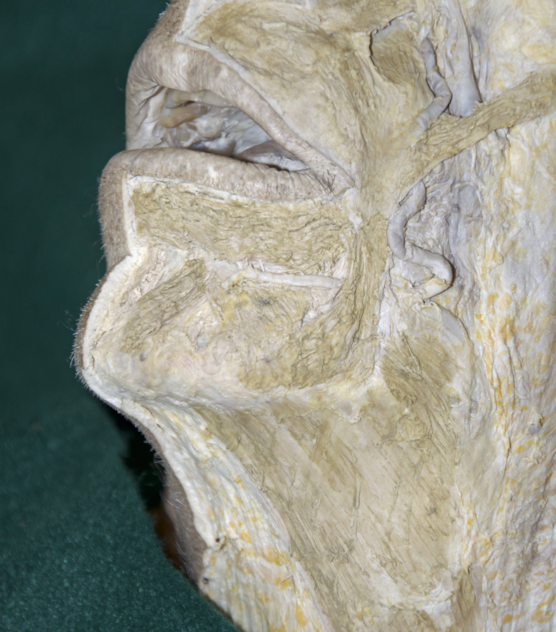
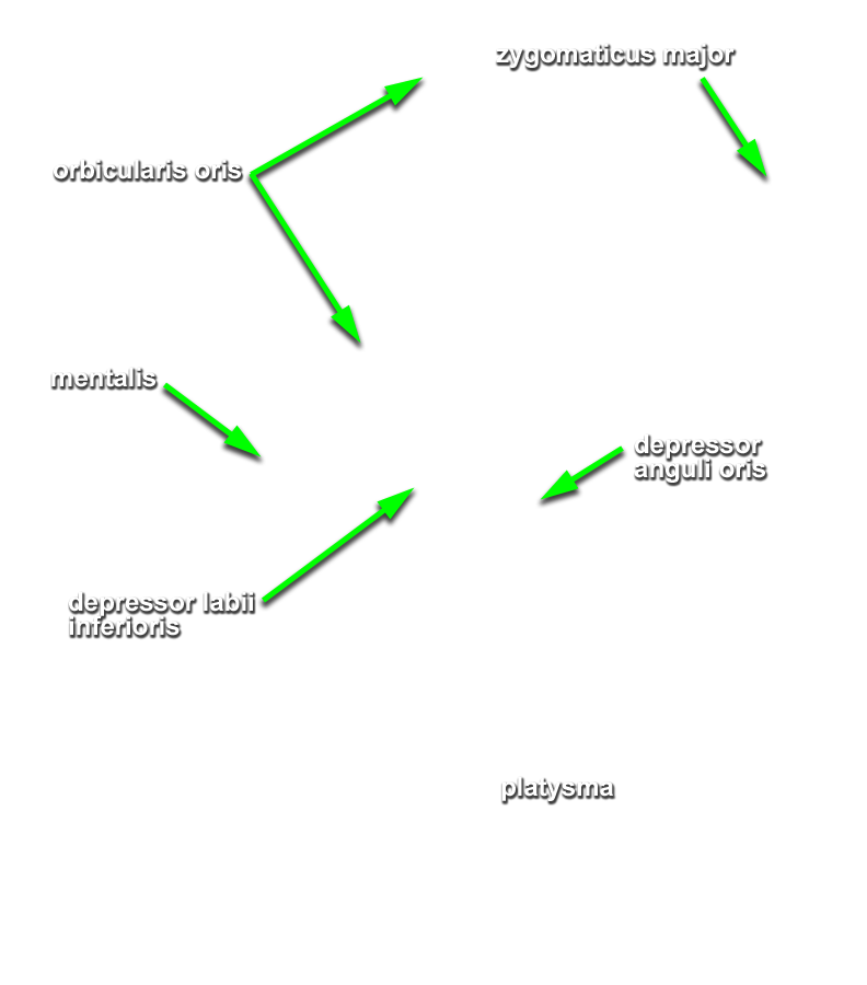
- Identify the
orbicularis oris,
levator anguli oris,
zygomaticus major,
depressor anguli oris and
levator labii superioris muscles. Attempt to identify the
alaeque nasi part of the levator labii superioris muscle, and the
depressor labii inferioris and
mentalis muscles. (G 7.12;N 25;Gl 38.1) Identify the
buccinator muscle. (G 7.16;N 48;Gl 40.21)
If you cannot locate the buccinator, then place the handle of your forceps in the oral cavity and push out on the cheek to bring the buccinator to a more superficial position. - Identify the platysma muscle where it attaches to the mandible. (G 7.13;N 25;Gl 38.1)