Bones
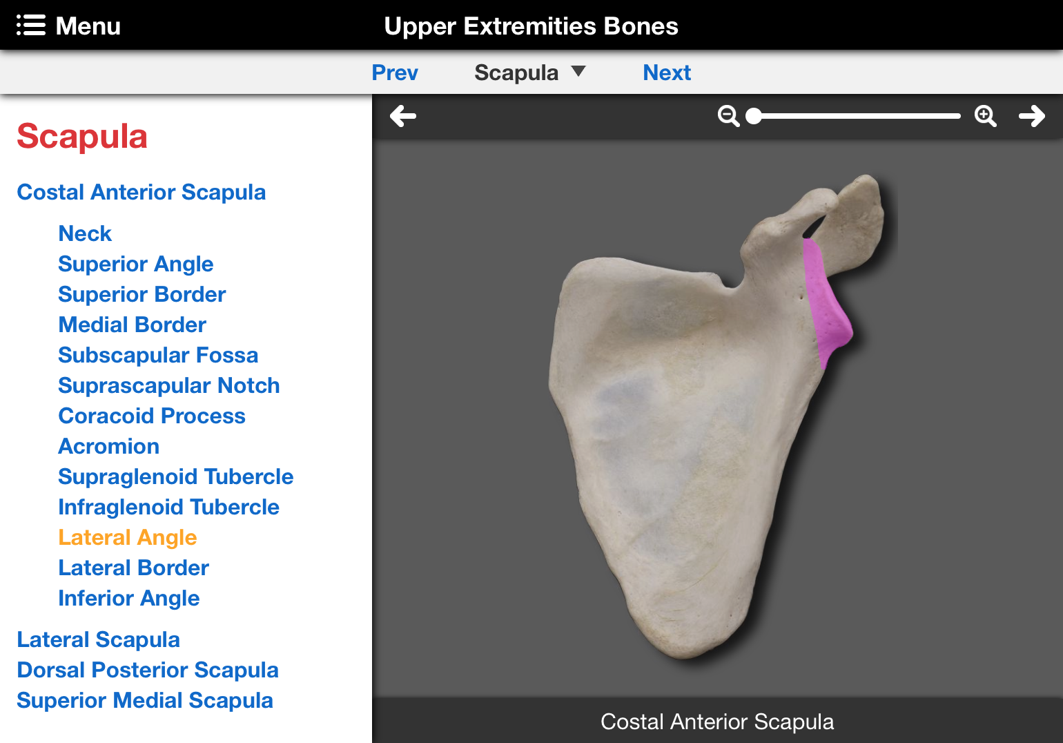
Individual bones (or collections) are listed in a centered drop-down menu. Each bone page presents one or more images of the bone along with a list of structures and landmarks relative to the bone or collection of bones being displayed.
- Click on a listed view to change the current view of the bone.
- Click on a listed landmark or structure to highlight it on the bone.
At the end of some bone lists there are interactive photographs for major ligament and muscle attachments. Major landmarks and their attachments, functions or details are listed on the left for each bone. Red structure names are hyperlinked to VH Dissector 3D and cross sectional views. Click on a red structure name to open the hyperlink.
Surface Anatomy
This section has two subsections with interactive photographs. Mouse over labels to highlight the corresponding structure or concept.
"Cutaneous Innervation" includes dermatomes and sensory cutaneous fields (for peripheral nerves). "Surface Landmarks" lists structures or landmarks, with red words hyperlinked to VH Dissector 3D and cross sectional views, that should be reviewed before starting the dissections for the region.
Movements
This section includes pairs of photographs demonstrating the movements occurring at the joints for that chapter. The joints (or their normally moving component) are listed in a centered drop-down menu. Click on the joint to bring up the movement(s) for that joint (listed under the joints). Click on each movement to bring up the photographs illustrating that movement.
Dissections
Each Dissection (Anterior and Medial Thigh, ...) includes "Overview", "Structures" and "Steps" sections.
Overview includes suggested readings from Moore's Clinically Oriented Anatomy and Essential Clinical Anatomy, and Drake's Gray's Anatomy for Students, and a summary description of the anatomical organization of the region to be dissected. The summary covers a description of the body surface, previews the skeleton and joints and presents the organization of the fascia or viscera of the region. A description of regional concepts including a summary of muscle function, innervation, blood supply and anatomical spaces/features concludes this section. Red words are hyperlinked to VH Dissector 3D and cross-sectional views. The Head and Neck chapter "Overviews" include interactive photographs illustrating the related muscles, nerves and arteries.
Structures is a summary list of anatomical structures to be identified in the dissected cadaver organized by systems. The "Structures" list may be useful for review prior to lab exams.
-
Steps are the numbered sequence of dissection instructions for an anatomical region or sub-region. Most Steps are subdivided into lettered sub-steps. University of Colorado students are required to identify bold (red, black or blue) structures. Red structures are hyperlinked to VH Dissector 3D and cross sectional views. Students are encouraged to dissect and identify all optional italicized structures. Most steps include photographs with labeled structures to the right of the dissection instructions. Camera icons ( ) in the text of the dissection instructions indicate that a relevant photograph is available. Click on a black camera icon (or color highlighted text immediately preceding it) to access the relevant photograph. The camera icon indicating the visible photograph is green. Red highlighted text immediately preceding a camera icon is a hyperlink to a relevant VH Dissector 3D and cross sectional view. Blue highlighted text immediately preceding a camera icon indicates relevant anatomy in the photograph. Alternatively, multiple photographs in a step can be accessed with forward/backward arrows above the photograph. Other controls above the photographs provide magnification and label control.
Atlas references are listed in parentheses for Grant's Atlas of Anatomy (G, 12th Ed.), Netter's Atlas of Human Anatomy (N, 5th Ed.) and Gilroy, MacPherson and Ross's Atlas of Anatomy (GI, 2nd Ed.). The dissection instruction steps include important relationships in yellow boxes and red words are hyperlinked to the VH Dissector as with the dissection instructions.
Observations
The Observations are designed for student groups that share (alternate) dissection laboratories and for students that observe previously dissected cadavers. Each "Observation" (Anterior and Medial Thigh, ...) includes the same "Overview" and "Structures" sections as their corresponding dissection and numbered "Steps" that match the numbering sequence of the dissection steps. Lettered sub-steps do not necessarily match the dissection sub-steps.
Observation "Steps" are the sequence of instructions to follow during the observation of a previously dissected anatomical region. Bold and colored text, photographs, atlas references and yellow "important relationship" boxes are used in the same manner as described in the "Dissections" section above.
Radiology
The Radiology section includes interactive radiographs (back, extremities, trunk, and head and neck), CT images (trunk, and head and neck), CT stacks (trunk) and MR images (head and neck). The interactive images are similar to the bone interactive images, except that there are no "show all" options. CT stacks highlight a single structure in a series (stack) of CT images. When viewing an imaging study, the corresponding 3D or cross sectional view in the VH Dissector is presented.
Summary
The Summary section includes an outline of anatomical structures and information based primarily on Gray's Anatomy (Susan Standring, Editor-in-Chief.; 39th Edition). The arteries, lymphatics, nervous system and veins are organized by branching pattern. The arteries include anastomoses, the lymphatics include afferent and efferent drainage, and the nerves include function(s). The bones, fascias, joints, ligaments, muscles and viscera (miscellaneous structures) are organized from superior to inferior along the head-trunk axis and proximal to distal along the extremities axes. Bones include the function (e.g., what traverses a landmark) and attachments at landmarks. Fascias include a summary of the details of the structure. The joints include classification and movements. Ligaments include their attachments and function. Muscles are further organized alphabetically by region and include attachments, function(s) and innervation. The viscera and miscellaneous structures include some details where appropriate.
The last category listed under Summary is "Relationships". The "Relationships" is a summary list of all the relationships (yellow boxes) included in all the dissections for that region.
Quizzes
The Quizzes are photographs from the dissection steps with pins marking most of the structures dissected and identified in the Cadaver Dissection Guide. The photographs are similar to the other "Interactive" photographs, except the name of the structure appears only when the cursor is over a red pin. Quizzes are accessed by clicking on the thumbnail image on the left side of the quizz page.