Place the cadaver in the prone position and carefully remove the skin surrounding the anus. Do not include any superficial fascia with the skin.
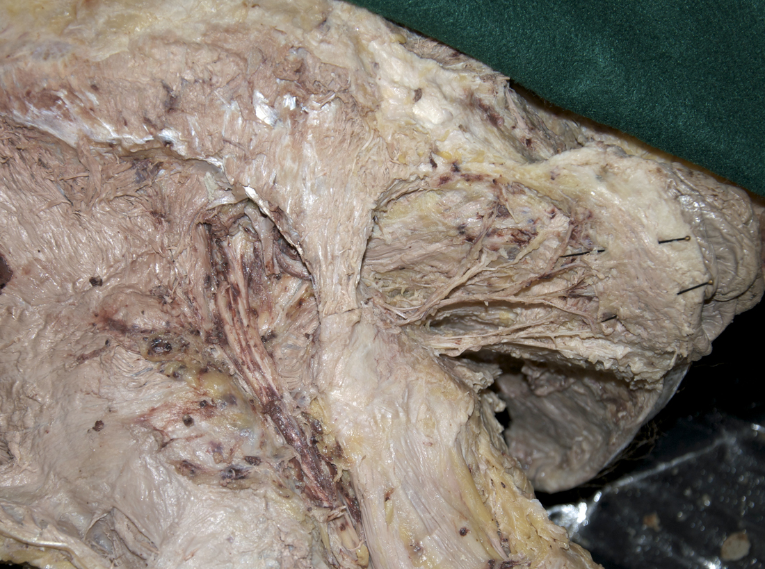
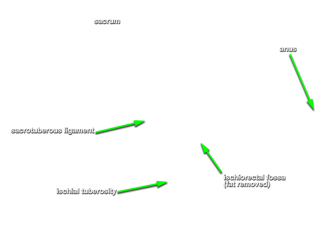
Identify and clean the nerves and vessels traversing the ischiorectal (ischioanal) fossa.
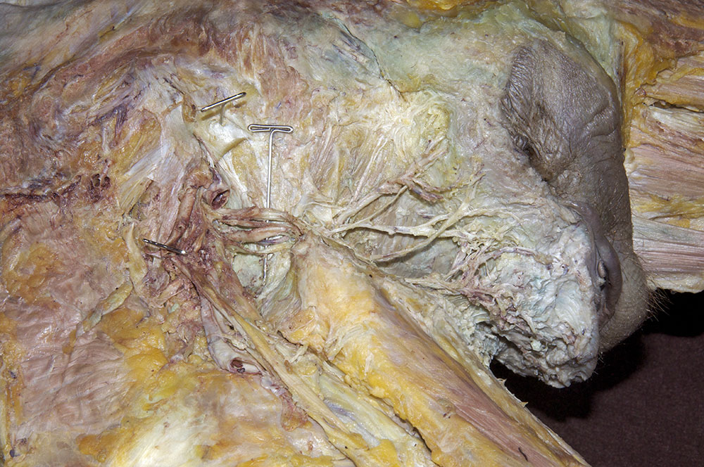
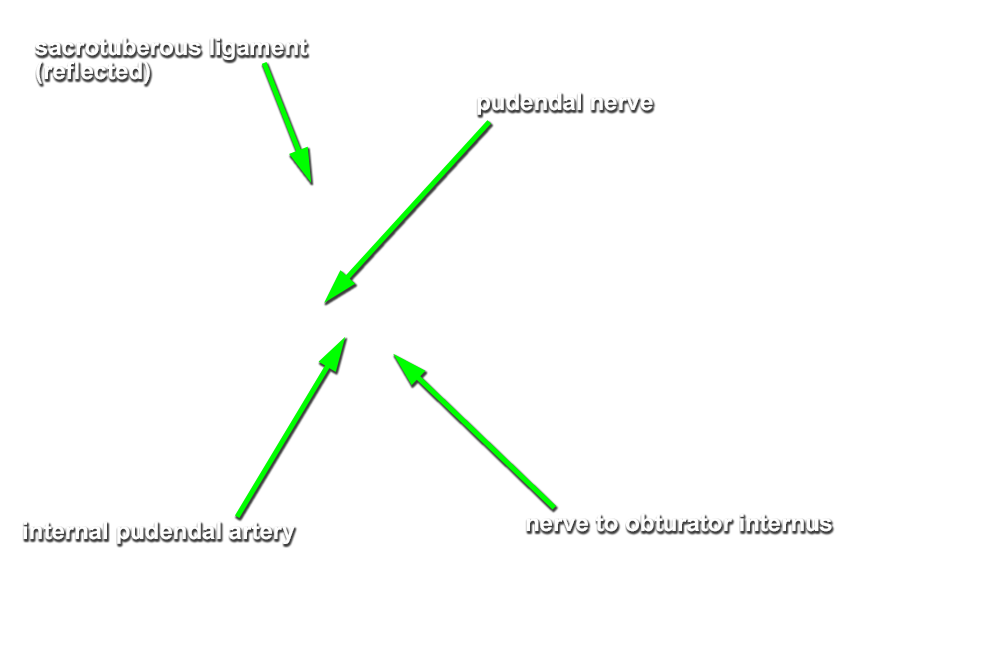
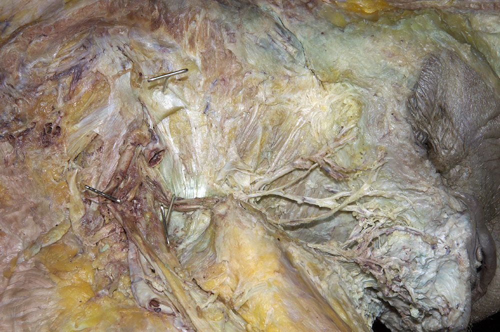
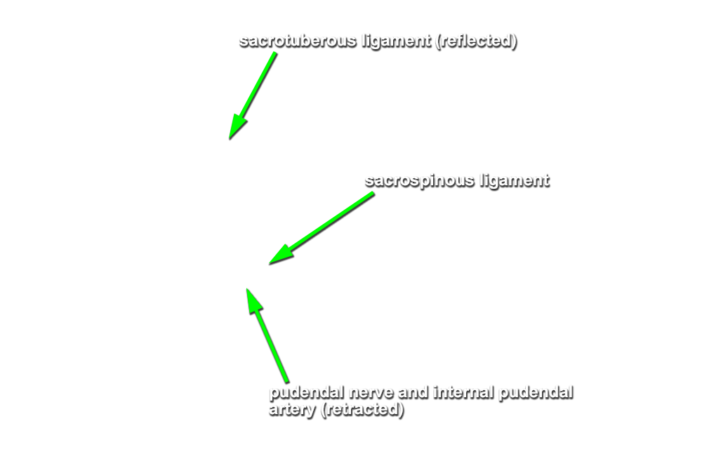
- Return to the sacrotuberous ligament. (G 6.31A;N 293 and 490;Gl 34.33) Blunt dissect the ligament away from the underlying nerves and vessels. Use your scissors to carefully cut the sacrotuberous ligament where it passes superficial to the sacrospinous ligament. (G 5.28C;N 393;Gl 26.8) Use a hemostat to reflect (inferior-lateral direction) and clamp the sacrotuberous ligament.
- Identify and clean the pudendal nerve and internal pudendal artery where these structures pass superficial to the sacrospinous ligament. You may remove the internal pudendal vein to clarify the dissection field.
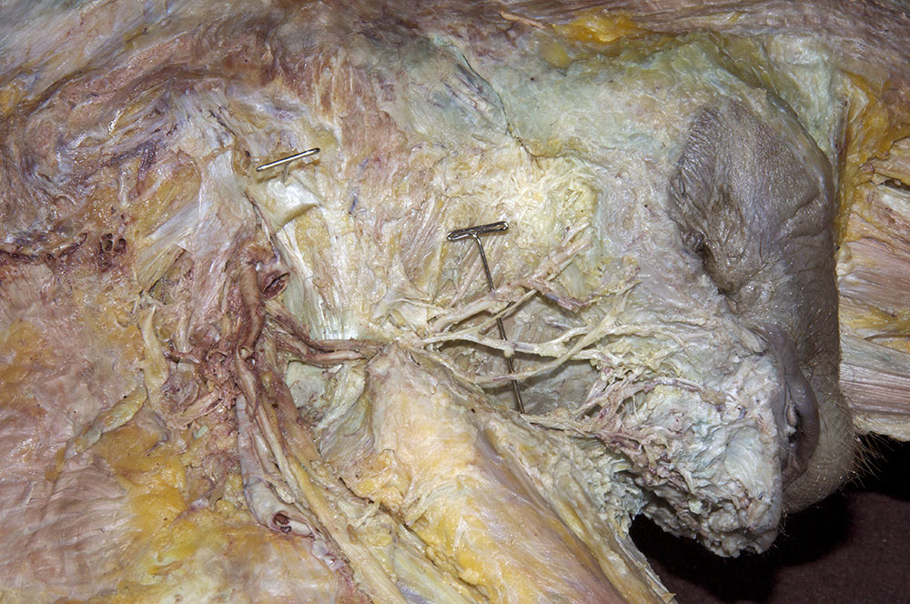
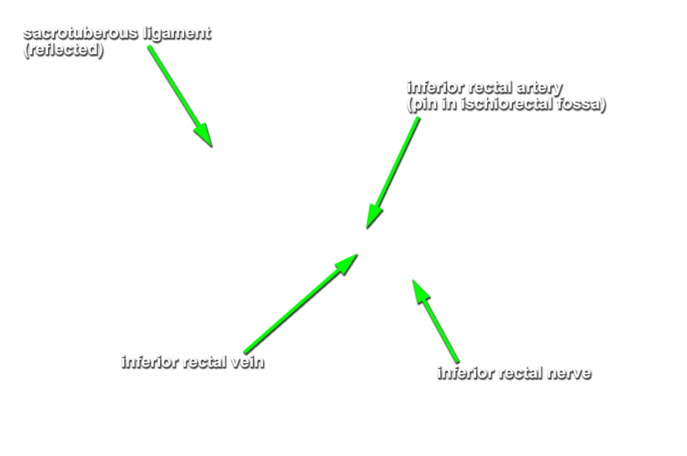
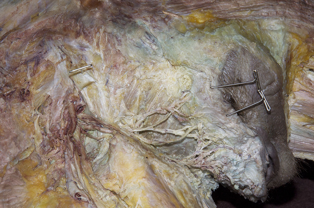
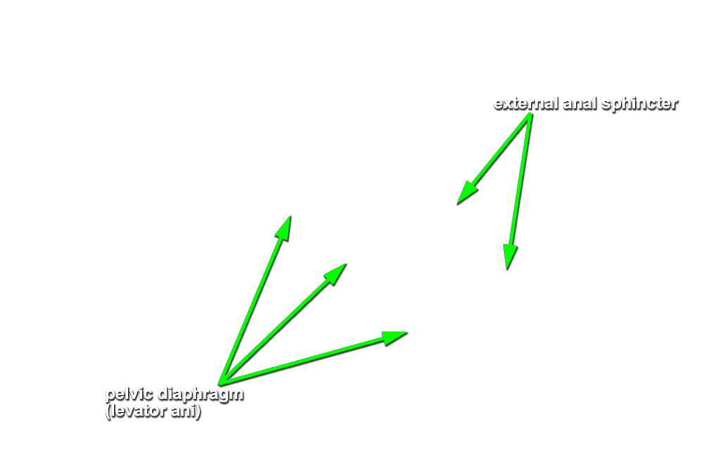
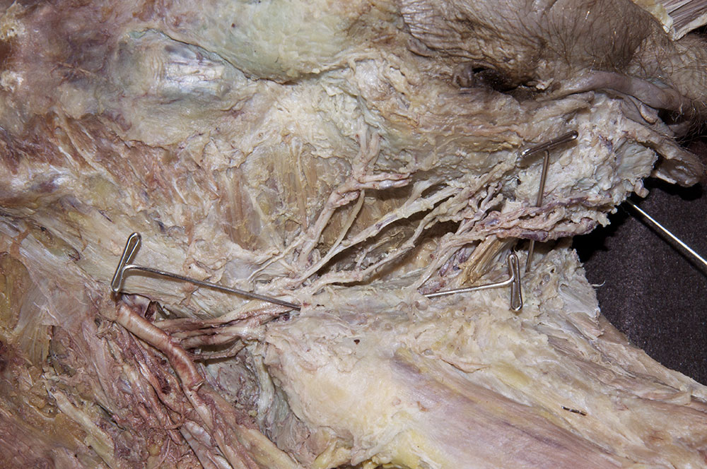
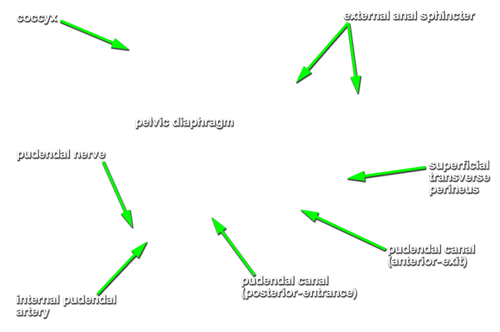
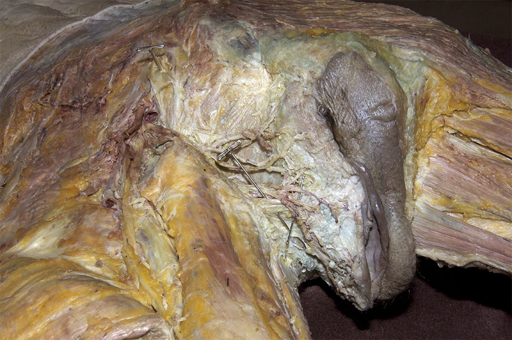
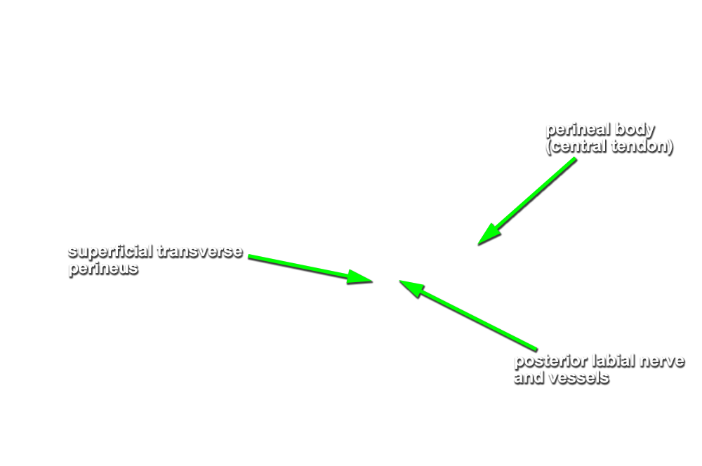
- Trace (blunt dissect) the branches of the pudendal nerve and internal pudendal artery into the ischiorectal fossa. Carefully remove the superficial fascia (fat). Identify the inferior rectal nerve and artery, and external anal sphinchter and levator ani muscle (pelvic diaphragm). (G 5.61;N 382 and 391;Gl 22.22)
- Use small scissors to open (blunt dissect) the fascia of the pudendal canal (tunnel in the obturator fascia) to expose the anterior continuation of the pudendal nerve and internal pudendal artery. (G 5.59A;N 391;Gl 22.22) Trace the pudendal nerve and internal pudendal artery branches as far anterior as possible (where they approach the labia majora).
Important Relationship
- The internal pudendal artery and vein, and the pudendal nerve pass posterior-lateral (superficial) to the sacrospinous ligament.