Use your hands to explore the pelvic cavity and its contents.
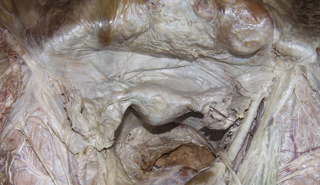
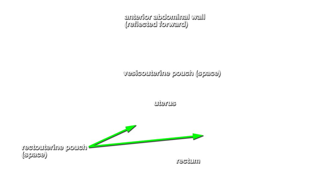
- Identify the rectum, rectouterine fold and pouch, and uterus. (G 5.30D;N 340;Gl 20.3)
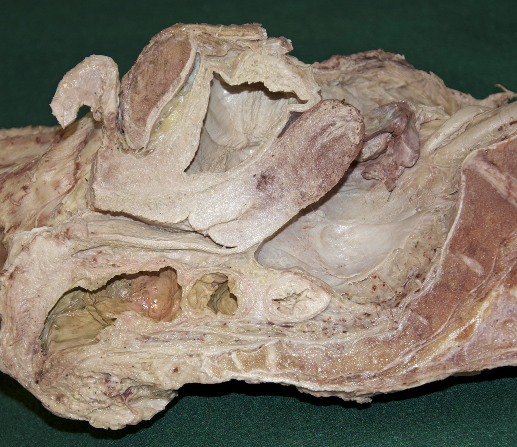
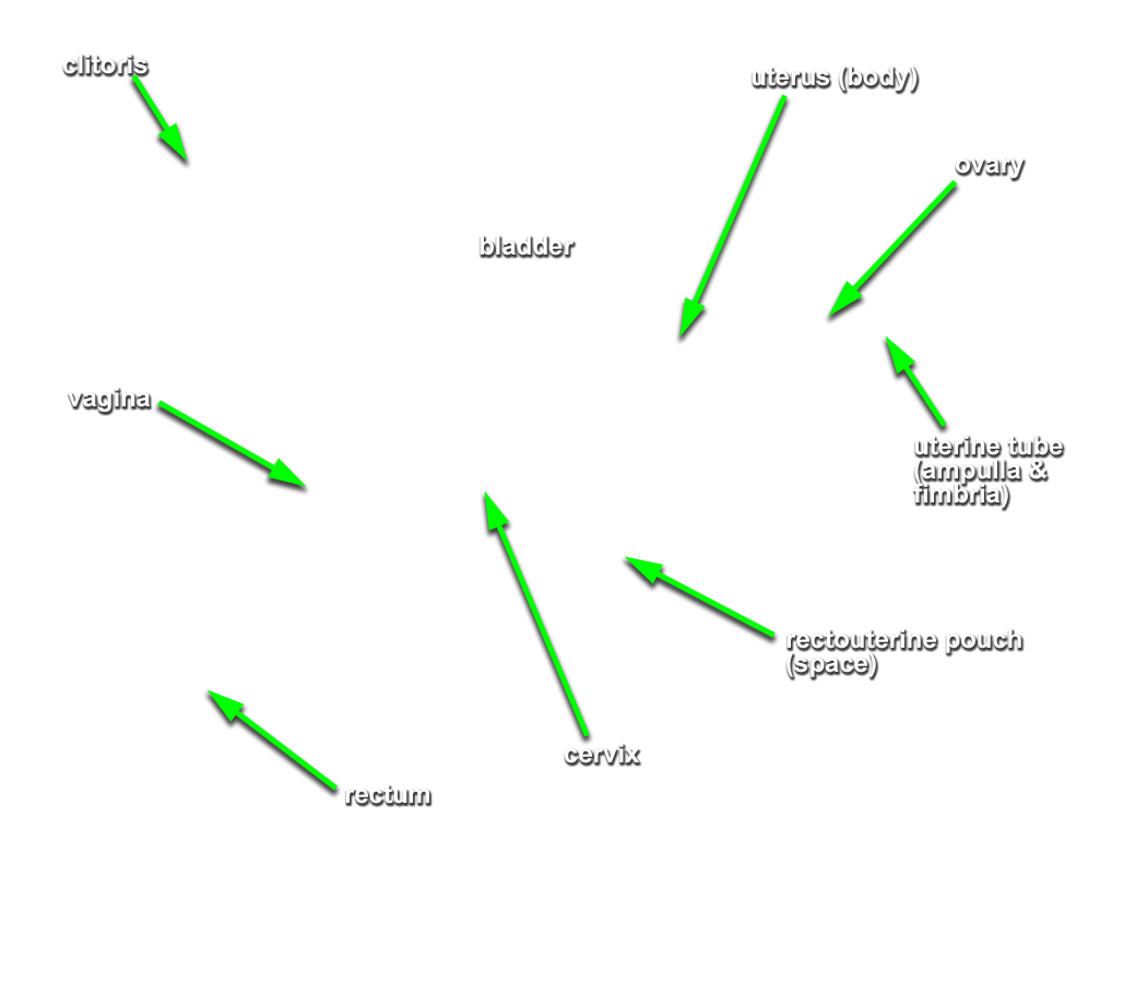
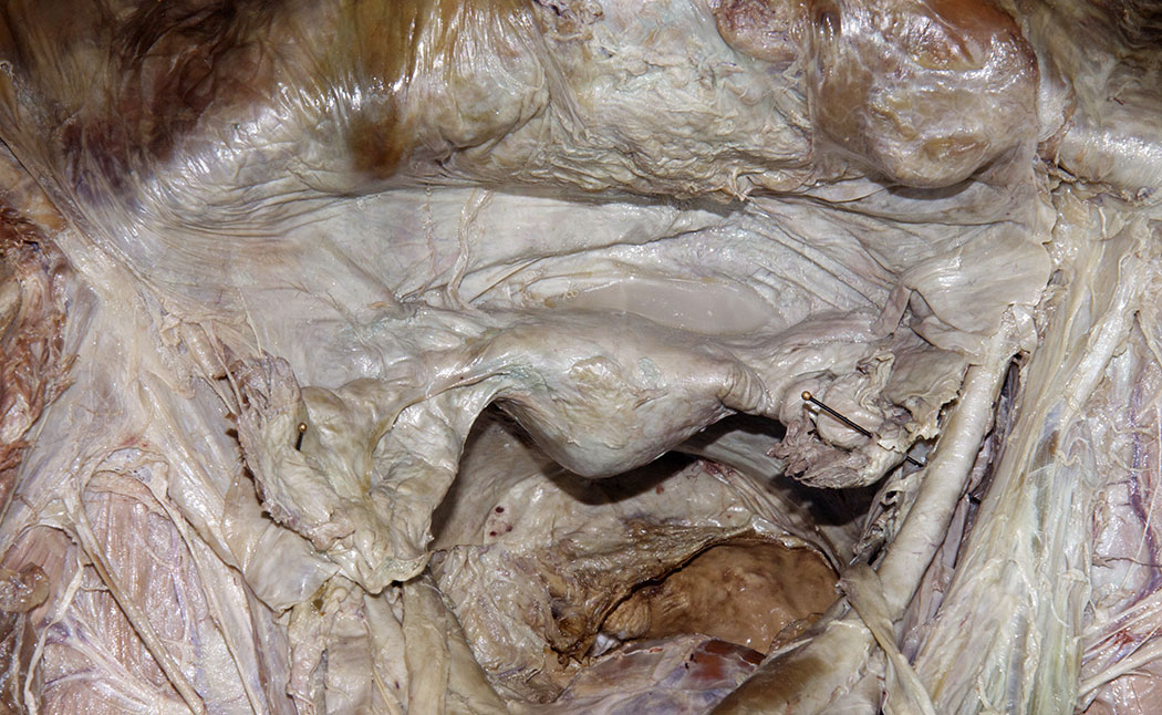
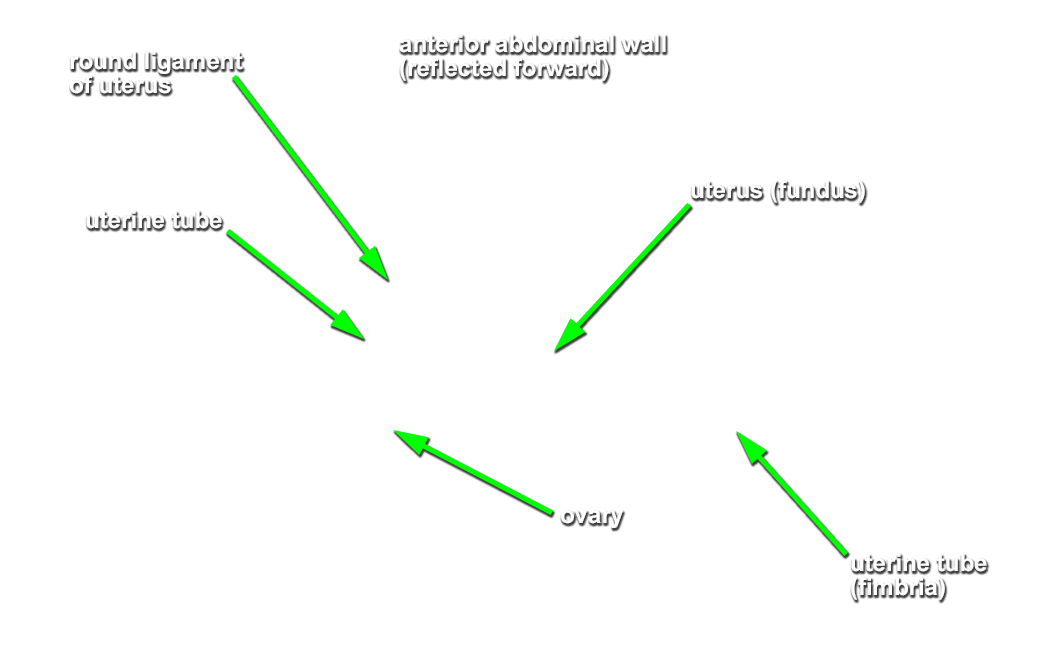
- Identify the uterine tube including the fimbriae. (G 5.33E;N 341;Gl 21.15) Place your index finger and thumb around the uterine tube. You are pinching the mesosalpinx.
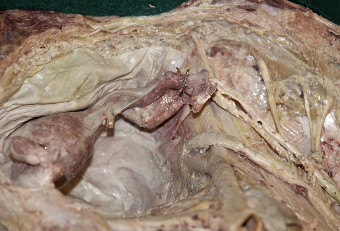
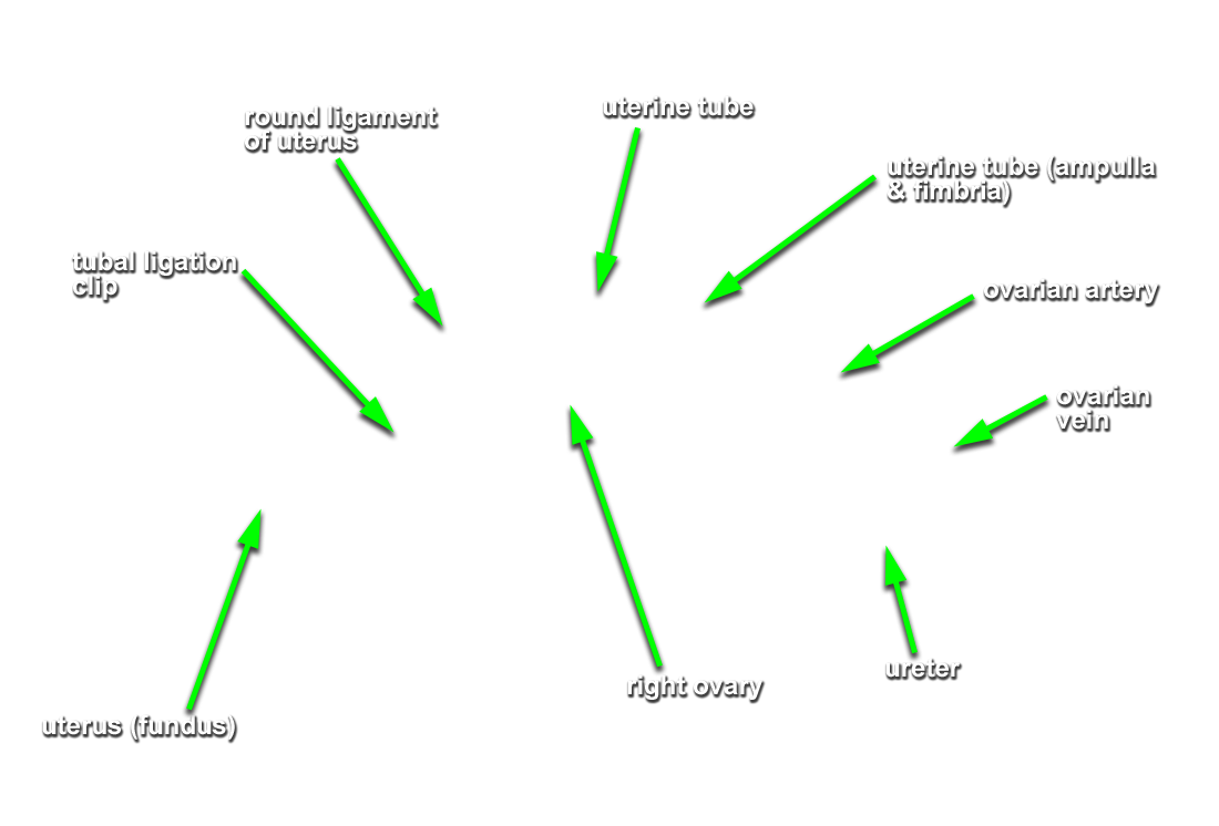
- Identify the ligament of the ovary, ovary and suspensory ligament of the ovary. Place your index finger and thumb around the ovary. You are pinching the mesovarium. On one side only ( ideally, the same side that you removed the psoas muscle), trace the ovarian artery and vein to the ovary by stripping away the peritoneum of the suspensory ligament.
- Identify the mesometrium and round ligament of the uterus. (G 5.30;N 353;Gl 20.3) On one side of the cadaver ( same side that you traced the ovarian artery and vein) strip away the peritoneum and trace the round ligament of the uterus to the deep inguinal ring. (G 5.30D;N 343;Gl 20.3)
- Identify the vesicouterine pouch and urinary bladder. (G 5.30D;N 343;Gl 20.3) Trace the ureter (same side that you previously traced the ovarian vessels and round ligament of the uterus) to the bladder (G 5.31;N 348;Gl 21.7) Attempt to identify the trigone, ureteric orifices and urethra. Attempt to identify the trigone and ureteric orifice.
Important Relationships
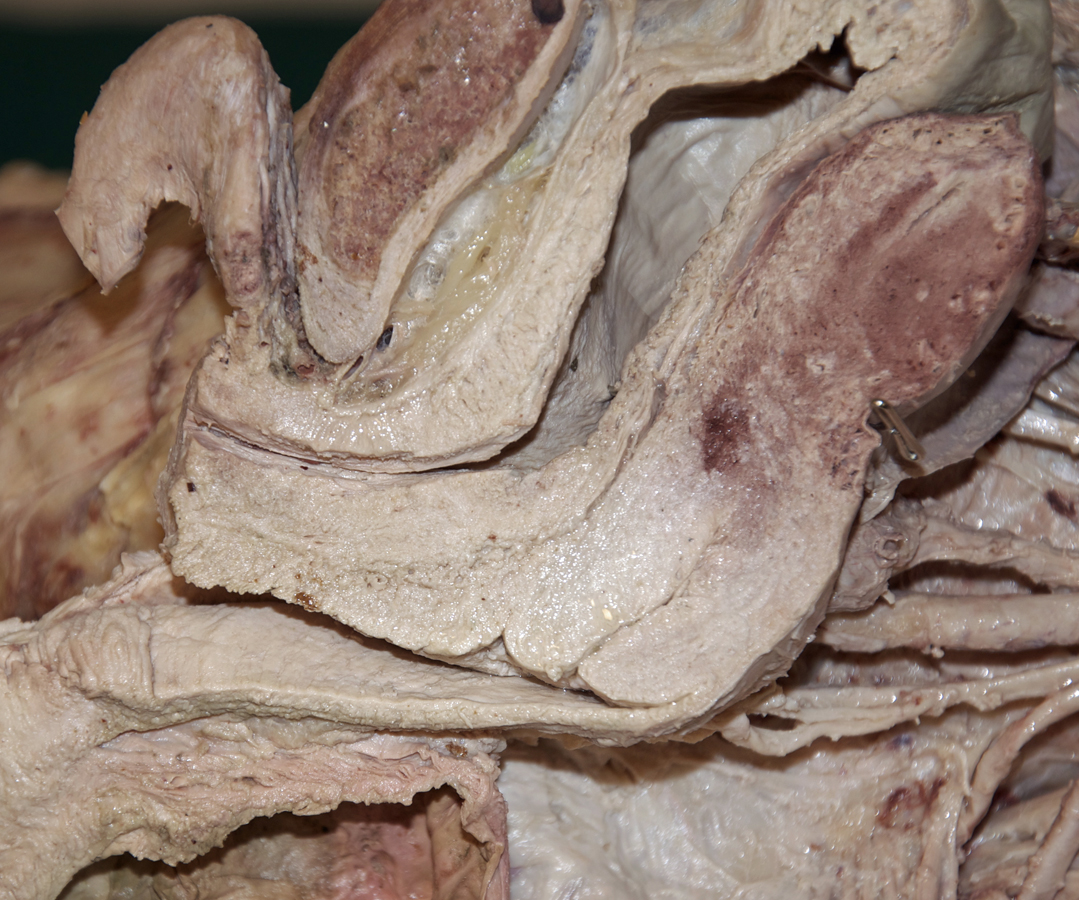
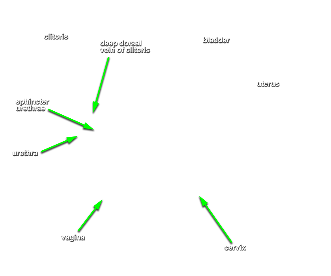
- The rectum is positioned posterior to the vagina and cervix and directly posterior to the rectouterine pouch.
- The uterus (body and fundus) is positioned posterior-superior to the bladder.
- The uterus is positioned directly anterior to the rectouterine pouch.
- The ovary is typically positioned posterior to the broad ligament of the uterus and posterior-inferior to the uterine tube.
- The urethra is positioned anterior to the vagina.