Identify the contents of the posterior mediastinum. (G 3.75;N 229;Gl 9.23)
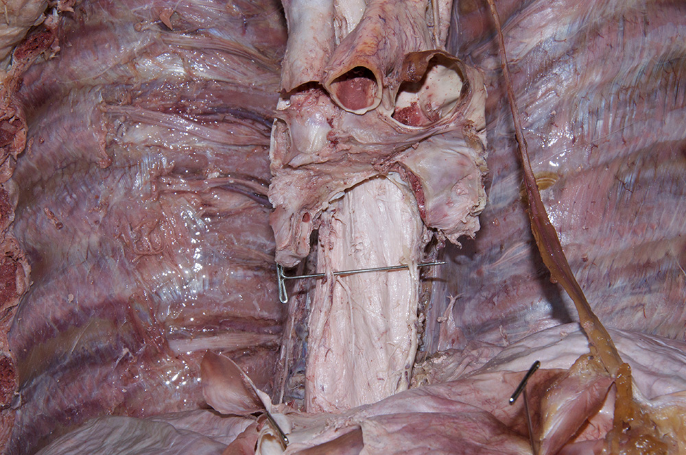
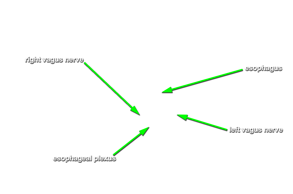
- Use scissors to dissect the posterior aspect of the pericardial sac from the esophagus and thoracic aorta, and reflect it towards the diaphragm. (G 3.46;N 229;Gl 8.11B) Trace the right and left vagus nerves to where they join the esophagus and contribute to the esophageal plexus.
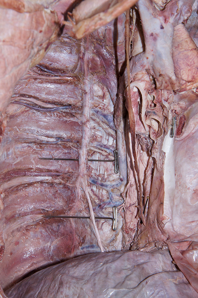
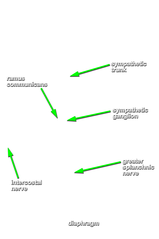
- Return to the sympathetic trunks. Identify the greater splanchnic nerves extending from the sympathetic trunks towards the midline. (G 3.75;N 227;Gl 9.3)
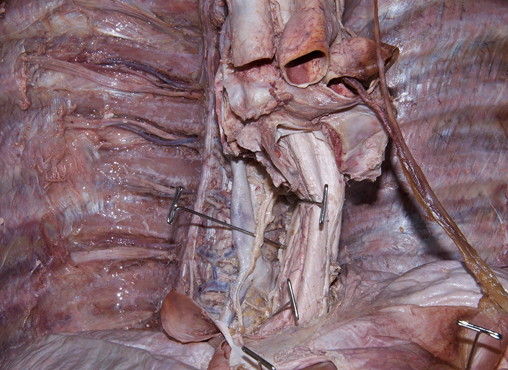
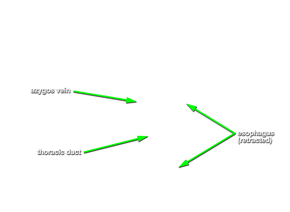
- Carefully explore (blunt dissection) the region posterior to the esophagus and medial to the descending aorta. Identify the thoracic duct. (G 3.70;N 235;Gl 8.7)
Important Relationships
- The descending (thoracic) aorta is positioned posterior to the pericardial sac.
- The descending (thoracic) aorta is positioned to the left (lateral) of the vertebral bodies.
- The inferior vena cava is positioned to the right of the descending aorta.
- The greater splanchnic nerve is positioned anterior - medial to the sympathetic chain.
- The esophagus is positioned posterior to the pericardial sac.
- Near the diaphragm, the esophagus is positioned anterior to the thoracic aorta.
- The thoracic duct is positioned directly anterior to the thoracic vertebral bodies.