Identify the structures associated with the posterior abdominal wall. (G 4.68A;N 308;Gl 16.17)
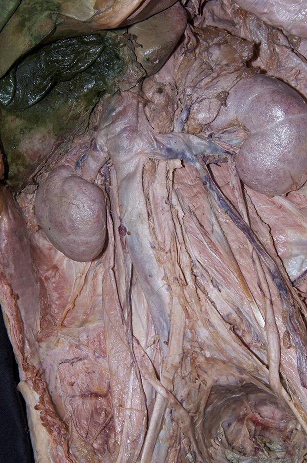
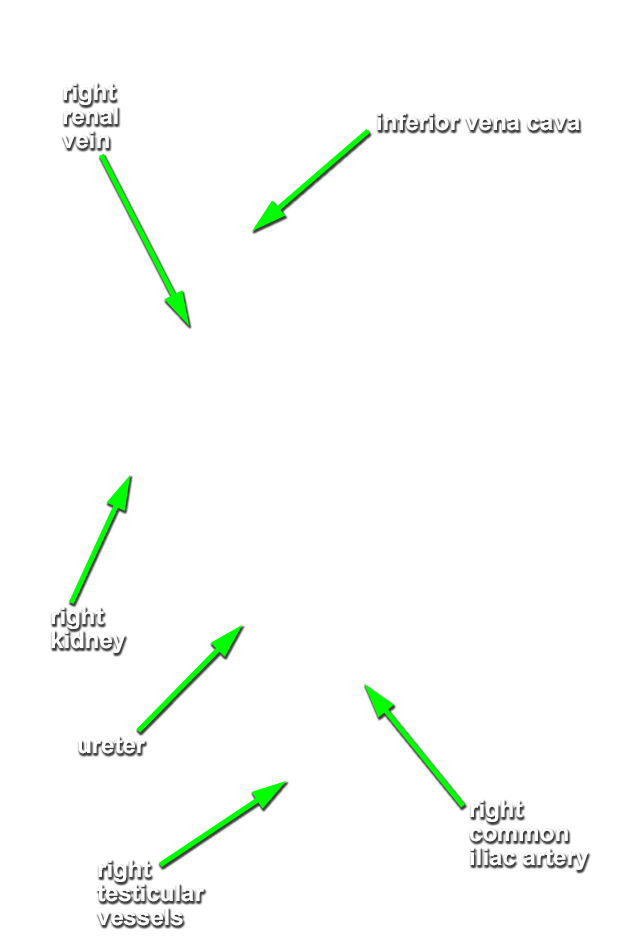
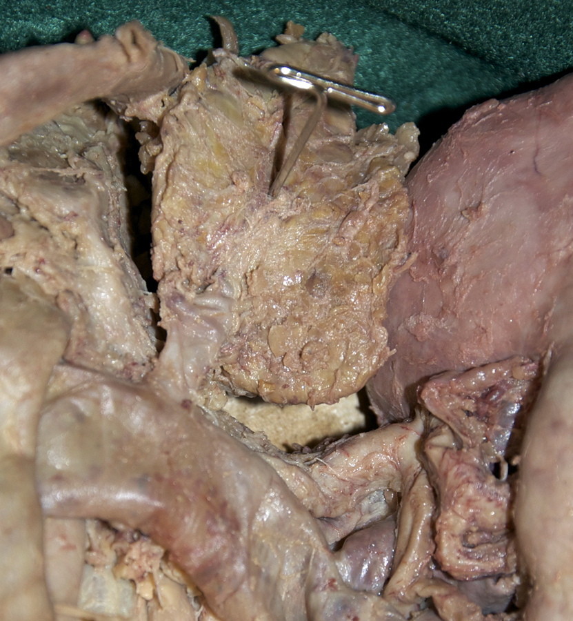
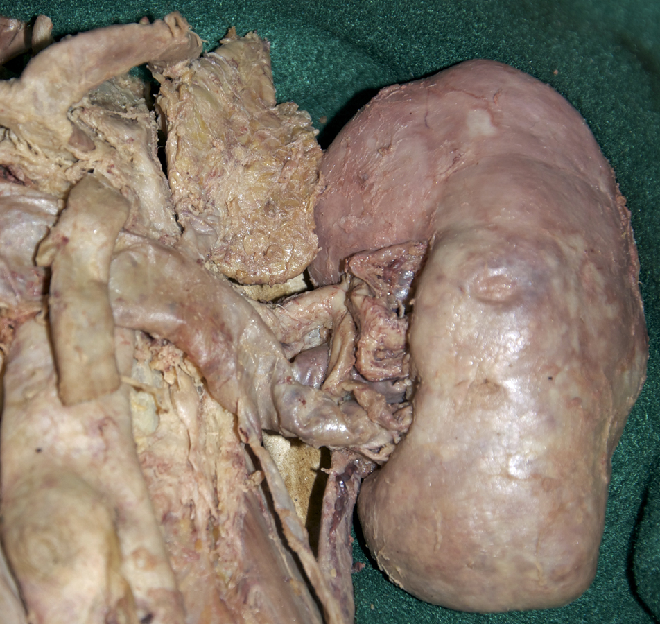
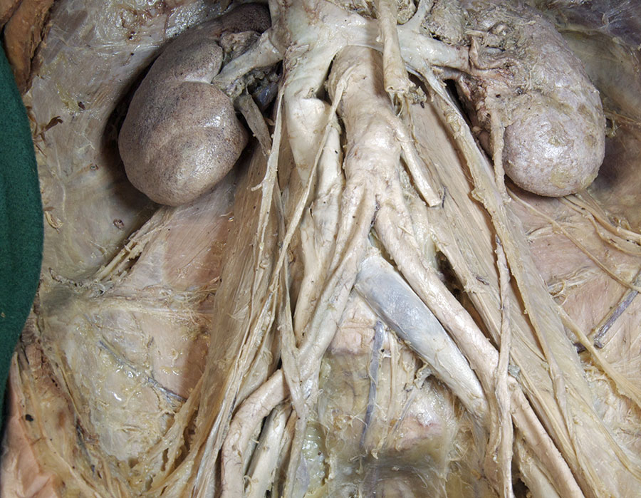
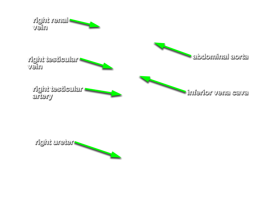
- Identify the kidneys and suprarenal glands. (G 4.69 and 4.76B;N 308;Gl 15.36) Identify the renal arteries and veins. Trace the renal arteries and veins to the abdominal aorta and inferior vena cava. Identify the left suprarenal vein where it drains into the left renal vein. Attempt to identify the inferior and middle suprarenal arteries arising from the renal artery and the aorta.
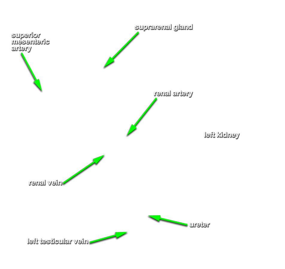
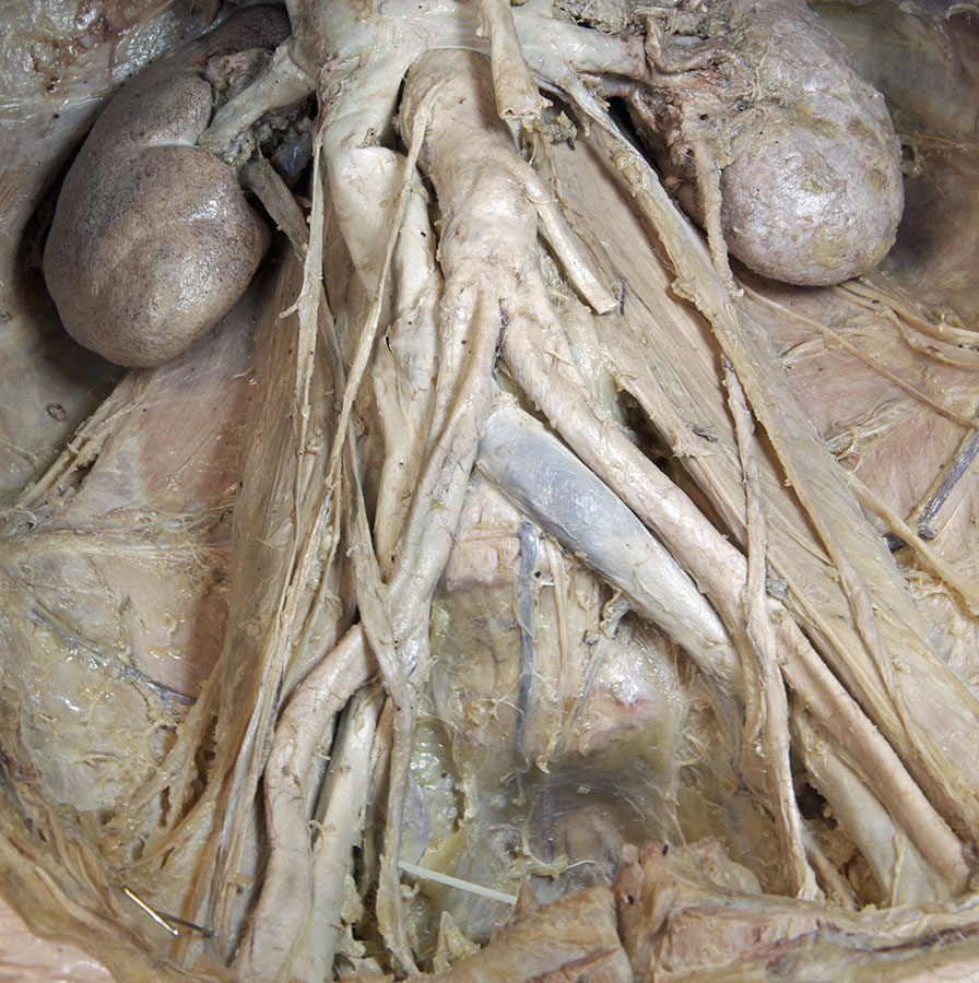
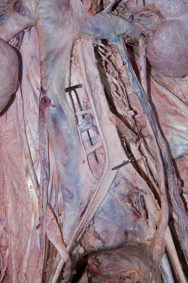
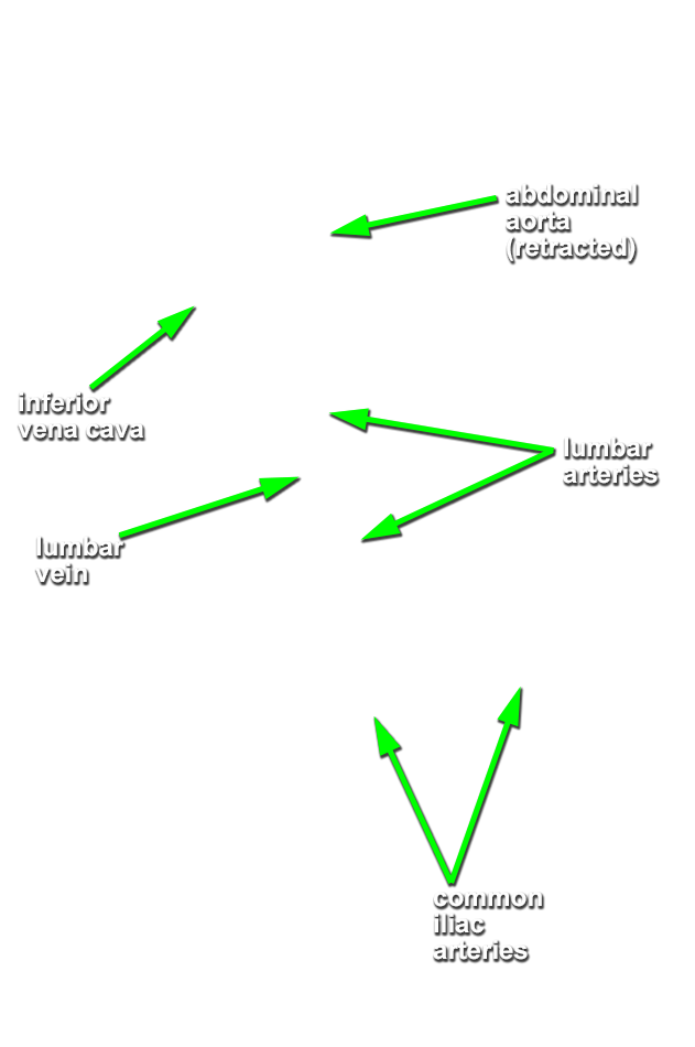
- Identify and trace the ureters to the level of the bifurcation of the abdominal aorta. (G 4.68A;N 308;Gl 16.17)
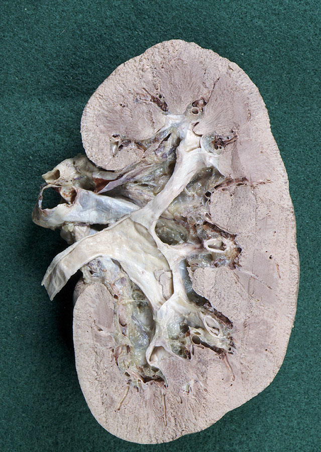
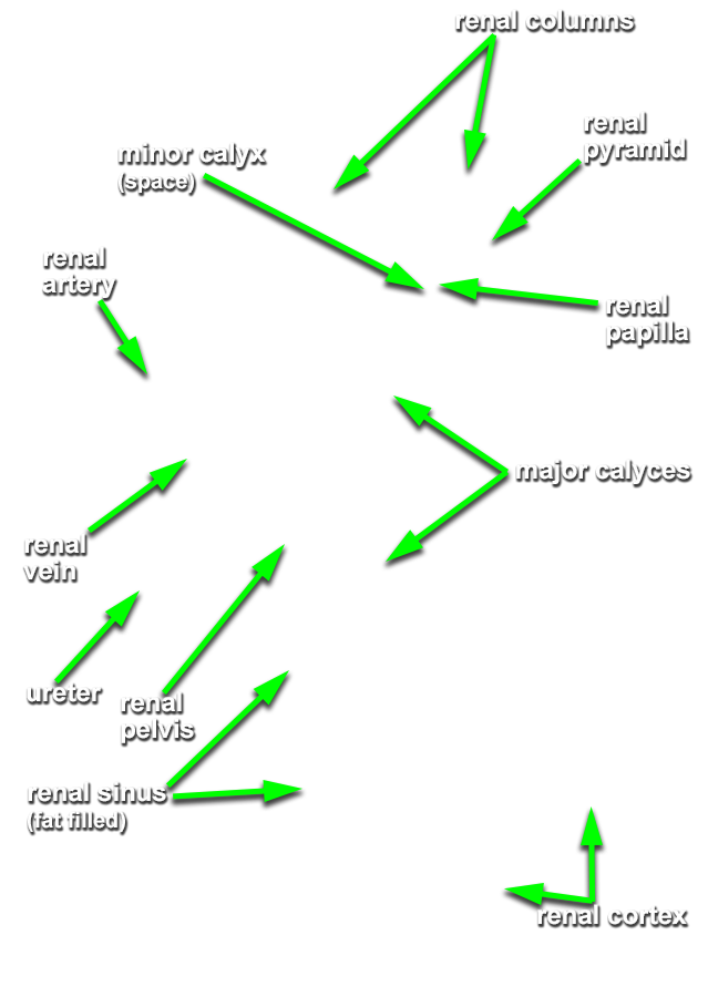
- Identify a renal column, renal pyramid, renal papilla, minor calyx, major calyx and renal pelvis in the bisected kidney.
- Identify the gonadal arteries and veins. (G 4.68;N 311;Gl 16.7)
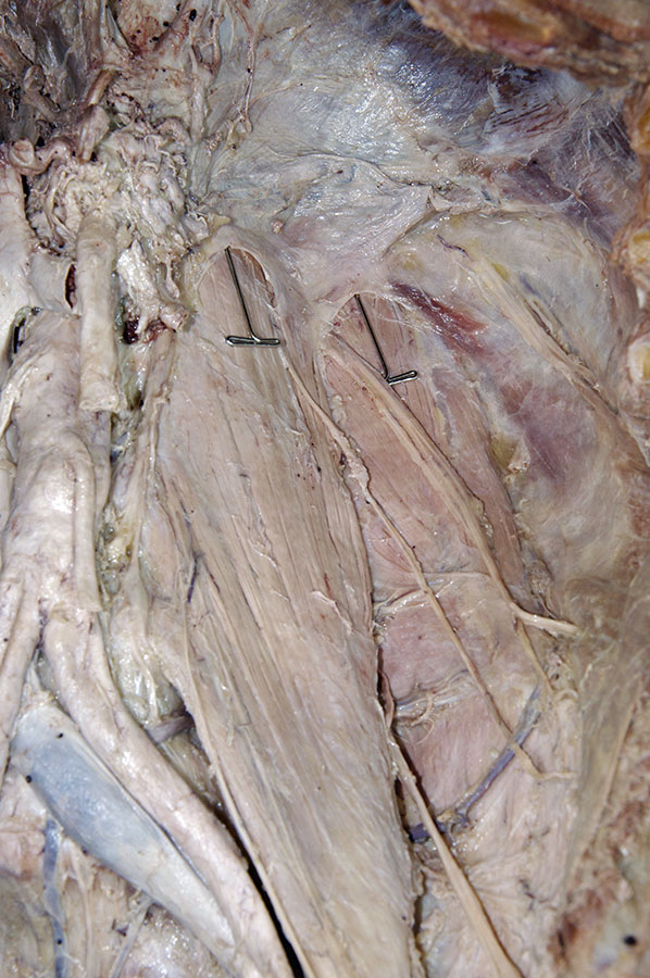
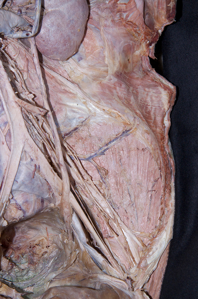
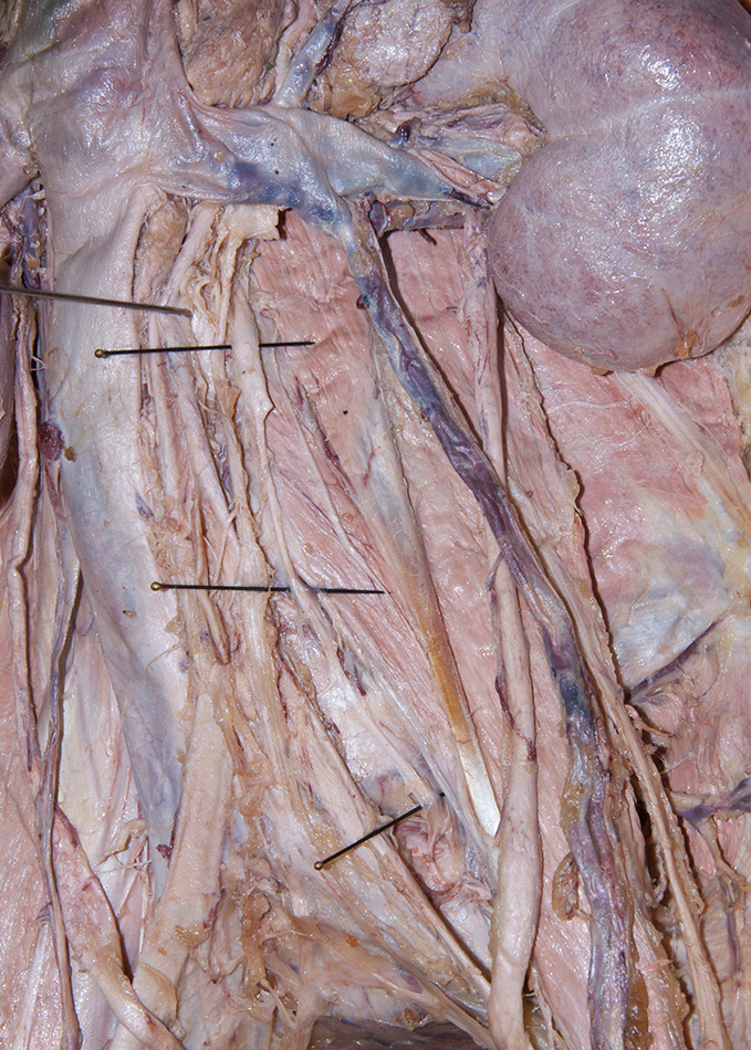
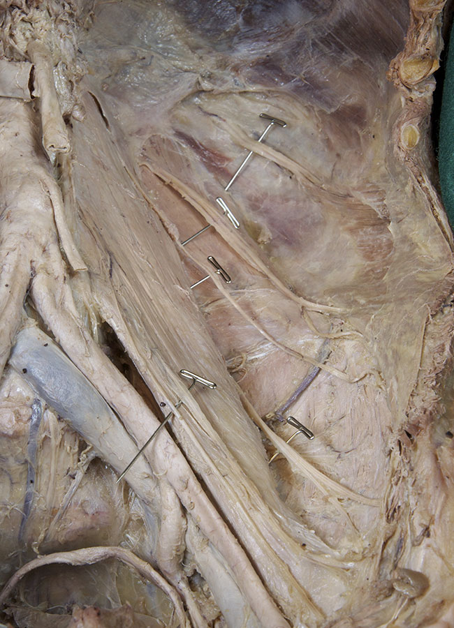
- Identify the psoas, quadratus lumborum and iliacus muscles. (G 4.77;N 258;Gl 13.5)
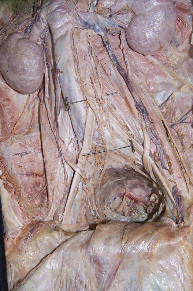
- Identify the common iliac arteries and veins. (G 4.68;N 259 and 260;Gl 16.16)
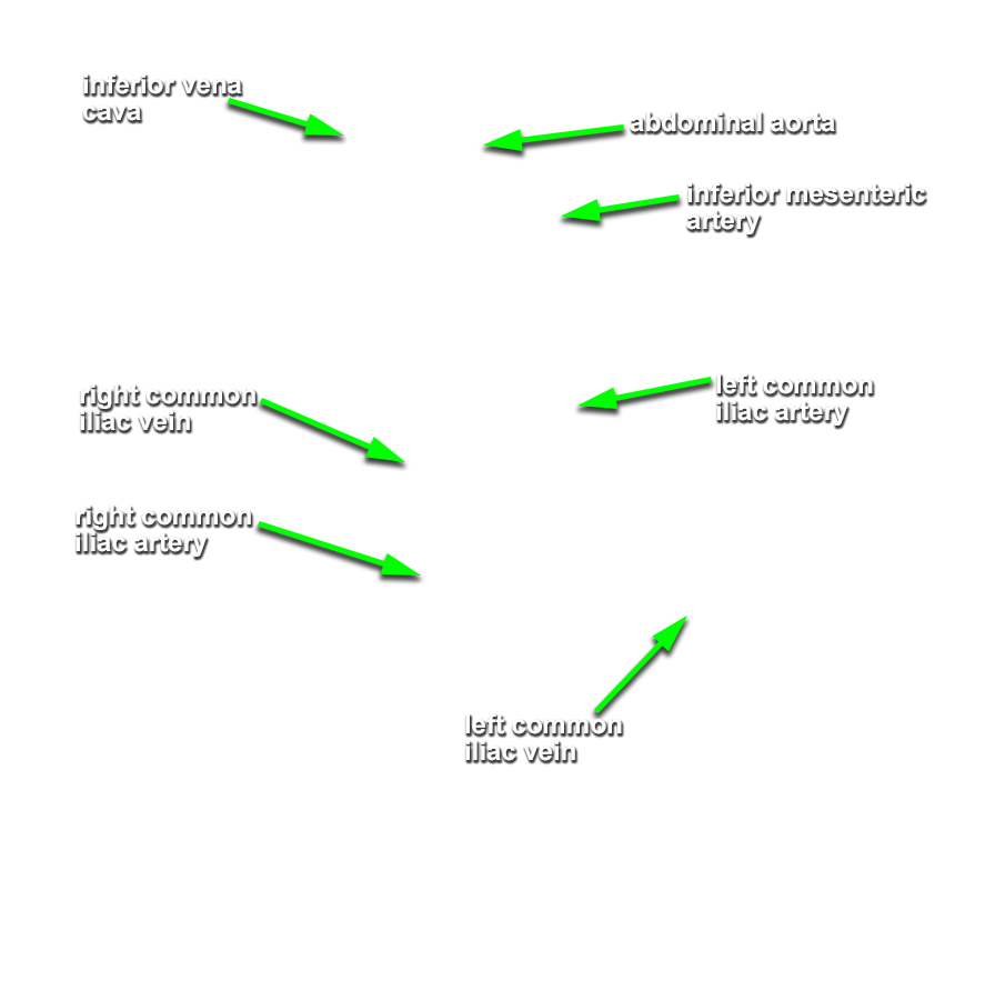
- Identify the left celiac ganglion (G 4.63A;N 262;Gl 16.40) and the sympathetic trunks. (G 4.77;N 262;Gl 16.40) Attempt to identify the hypogastric plexus.
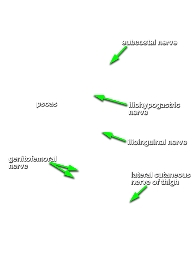
- Identify the subcostal, iliohypogastric, and ilioinguinal nerves, the lateral cutaneous nerve of the thigh, the femoral, genitofemoral, and obturator nerves and the lumbosacral trunk. (G 4.78;N 262;Gl 16.37) Identify a lumbar artery and vein. (G 4.80A;N 259;Gl 16.7)
Important Relationships
- The kidney is positioned anterior - lateral to the psoas muscle and anterior to both the quadratus lumborum and transversus abdominis muscles.
- The right suprarenal gland is positioned superior to the right kidney.
- The left suprarenal gland is positioned superior-medial to the left kidney.
- The right renal artery passes directly posterior to the inferior vena cava.
- The left renal vein passes anterior to the abdominal aorta and posterior to the superior mesenteric artery.
- The right ureter passes anterior to the psoas muscle.
- The right testicular artery passes directly anterior to the inferior vena cava, the right ureter, and the psoas muscle.
- The right common iliac artery passes directly anterior to the left common iliac vein.