Reading
- p. 979 (Sympathetic ...) - 981 (Lymphatics)
- p. 1012 - 1018 (Viscera ...)
- p. 1051 (Lymphatics ...) - 1052
- p. 586 (Pretracheal)
- p. 604 - 605
Body Surface
The primary surface landmarks of the deep neck are associated with the external ear (external auditory meatus) and temporal bone (mastoid process). Branches of the cervical plexus (lesser occipital, great auricular and transverse cervical nerves) innervate the skin overlying the deep neck structures.
Skeleton and Joints
The bones associated with the deep neck are the occipital and temporal bones and cervical vertebrae, including the atlas and axis. The cervical spine is supported by the anterior longitudinal, posterior longitudinal, ligamenta flava, intertransverse, interspinous and supraspinous ligaments. There are facet (zygapophyseal) joints between adjacent superior and inferior articular processes, and symphases (intervertebral discs) between the adjacent vertebral bodies. The cervical spine moves in flexion, extension, lateral flexion and rotation. The atlantoaxial and atlanto-occipital joints are supported by fibrous joint capsules, the cruciate and alar ligaments, and the tectorial and anterior and posterior atlanto-occipital membranes. The two atlanto-occipital joints (superior articular processes and occipital condyles) form an ellipsoidal articulation allowing flexion and extension and slight lateral flexion of the head. The median atlantoaxial joint is a pivot articulation, and the two lateral joints are gliding articulations. Together they allow for rotation of the head.
Organization
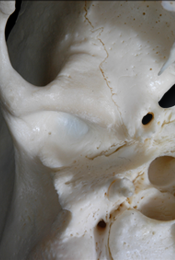
The deep neck region includes the parotid bed, the cranial base medial to the parotid bed, the prevertebral muscles and fascia, and the atlantoaxial and atlanto-occipital joints. The partotid bed is the space between the ramus of the mandible, styloid process and mastoid process. The cranial base medial to the partotid bed houses the openings of the hypoglossal canal, jugular foramen, carotid canal and stlyomastoid foramen. The prevertebral fascia is thick and prominent where it covers the anterior vertebral muscles and is thin and non-distinct deep to the trapezius. It covers the cervical ventral rami and is continuous with the axillary sheath. The carotid sheath is a fascial tube surrounding the common and internal carotid arteries, the internal jugular vein, the vagus nerve and portions of the ansa cervicalis. It is positioned anterior to the prevertebral muscles and deep to the sternocleidomastoid.
Muscles
The prevertebral muscles (longus capitis, longus colli, rectus capitis anterior and lateralis, and scalenus anterior, middle and posterior) function to flex, laterally flex and rotate the head and neck.
Innervation
Branches of the ventral rami of cervical spinal nerves innervate (motor, sensory and postganglionic sympathetic) the prevertebral muscles.
Blood Supply
Branches of the vertebral arteries and thyrocervical trunks supply, and tributaries of the retromandibular, external jugular, internal jugular, vertebral and left brachiocephalic veins drain the deep neck structures.
