Reading
- p. 822 - 855
- p. 880 (Orbital Septum) - 902 (Ear)
- p. 908 (Cranial ...) - 916 (Temporal Region)
- p. 499 (Cranial ...) - 514 (Face)
- p. 524 (Orbit) - 539 (Temporal ...)
- p. 578 - 581
Body Surface
The primary surface landmarks of the cranial cavity and orbit are associated with the eye (supraorbital margin, infraorbital margin, palpebral fissure, medial and lateral commissures, and lacrimal caruncle, lake and punctum). Branches of the trigeminal (nasociliary, supratrochlear, supraorbital, lacrimal, infraorbital and auriculotemporal), lesser occipital and greater occipital nerves innervate the skin overlying the cranial cavity and orbit.
Skeleton and Joints
The bones associated with the cranial cavity are the frontal, ethmoid, sphenoid, parietal, temporal and occipital. The frontal, sphenoid, zygomatic, maxilla, lacrimal, ethmoid and palatine bones contribute to the walls of the orbit. There are sutures (fibrous joints) between the adjacent bones.
Organization
In this dissection exercise you study the cranial cavity, gross brain and orbit. The brain is subdivided into the cerebrum, brainstem and cerebellum. The cerebrum has two massive hemispheres each with four (frontal, parietal, occipital and temporal) lobes, and a more deeply placed diencephalon. One cranial nerve (olfactory) is associated with the cerebral hemispheres and one cranial nerve (optic) arises from the diencephalon. The cerebellum has two hemispheres and a midline vermis. The brainstem is subdivided into the midbrain, pons and medulla, and the remaining crainial nerves arise from the brainstem.
The cranial cavity is subdivided into three regions, the anterior, middle and posterior cranial fossae. The anterior cranial fossa is positioned superior to the orbits and nasal cavity. The anterior fossa houses the poles of the frontal lobes. The middle cranial fossa is associated with the sphenoid bone and extends from its lesser wings anteriorly to the petrous portion of the temporal bone posteriorly. Along the midline, it includes the sella turcica. The poles of the temporal lobes rest in the floor of the middle cranial fossa, and cranial nerves II (optic), III (oculomotor), IV (trochlear), V (trigeminal) and VI (abducens) exit the cranial cavity from this fossa. The posterior cranial fossa extends from the petrous portion of the temporal bone to the foramen magnum. It houses the brainstem and cerebellum. Cranial nerves VII (facial), VIII (vestibulocochlear), IX (glossopharyngeal), X (vagus), XI (accessory) and XII (hypoglossal) exit the skull from the posterior cranial fossa.
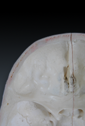
Three concentric membranes, the meninges, surround the brain. The outer most membrane, the dura mater, lines (periosteum) the bones of the cranial cavity. Along the midline, the dura is reflected inferiorly forming the sickle-shaped falx cerebri. The falx cerebri separates the two cerebral hemespheres and extends from the crista galli to the internal occipital protuberance. The posterior aspect of the falx cerebri is broad and splits laterally to form the tentorium cerebelli. Its lateral margin attaches to the occipital bone and petrous portion of the temporal bone. It extends anteriorly to attach to the clinoid processes of the sphenoid (sella turcica) bone. The tentorium cerebelli separates the occipital lobes from the cerebellum. The medial free margin of the tentorium cerebelli forms a notch or apperature surrounding the midbrain. A small extension of dura, the diaphragma sella forms the roof of the sella turcica. The dural venous sinuses are venous channels located in splits within the dura mater.
The orbit houses the eye, the muscles (extraocular) that move the eye, the lacrimal gland, the nerves and vessels that supply the eye, and nerves and vessels that traverse the orbit to reach the nasal cavity or face. The orbit has a weak fibrous sheath, the orbital septum, that extends from the periosteum of the orbital margin to the tarsal plates of the upper and lower eyelids. There are medial and lateral palpebral ligaments attached to the edges of the tarsal plates. Abundant orbital fat fills the spaces between the eye and the muscles, nerves and vessels.
Muscles
Six extraocular muscles (superior rectus, medial rectus, inferior rectus, lateral rectus, superior oblique and inferior oblique) function to move (elevation, depression, abduction, adduction, intorsion and extorsion) the eye. The levator palpebrae superioris functions to open and the orbicularis oculi functions to close the upper lid.
Innervation
The optic nerve (vision) and branches of the nasociliary nerve (ophthalmic division of the trigeminal nerve) innervate (sensory) the eye. Branches of the oculomotor, trochlear and abducens nerves innervate (sensory, motor and postganglionic sympathetic) the extraocular muscles and the levator palpebral superioris. Intraocular smooth muscle (iris and ciliary body) is innervated by postganglionic sympathetic axons from the superior cervical ganglion and postganglionic parasympathetic axons from the ciliary ganglion. The lacrimal gand is innervated by postganglionic sympathetic axons from the superior cervical ganglion and postganglionic parasympathetic axons from the pterygopalatine ganglion.
Blood Supply
Branches of the middle meningeal artery are the primary supply of the dura mater lining the cranial cavity. Branches of the internal carotid, vertebral and basilar arteries supply the brain. Tribuaries of the dural venous sinuses drain the brain. The dural venous sinuses are tributaries of the internal jugular vein. Branches of the ophthalmic artery supply the structures of the orbit and tributaries of the superior and inferior ophthalmic veins drain the same structures. The superior and inferior ophthalmic veins are tributaries of the cavernous sinus.