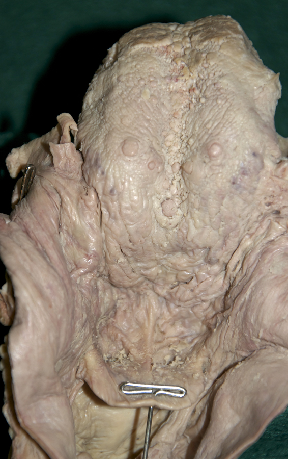
Identify the internal surface features and structures associated with the laryngeal pharynx and larynx.
- Identify the
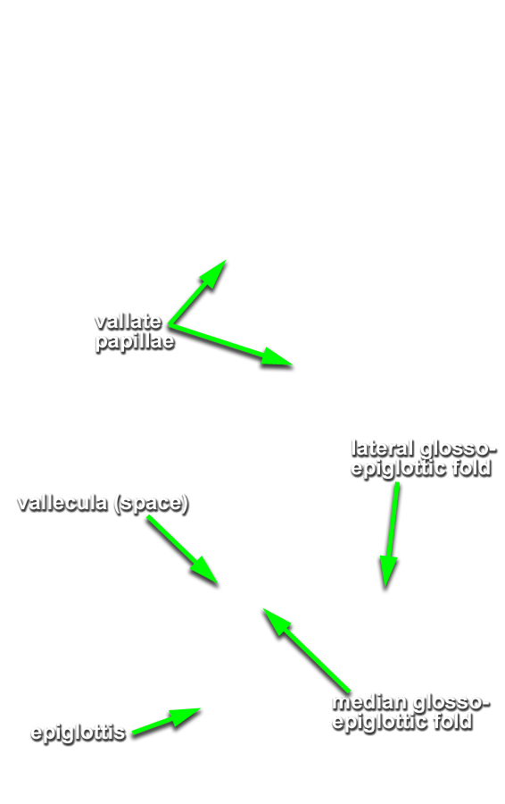
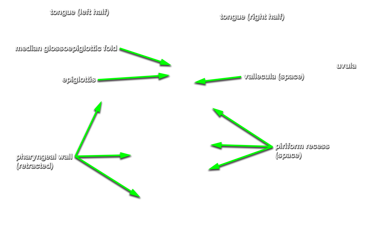
median and
lateral glossoepiglottic folds,
vallecula,
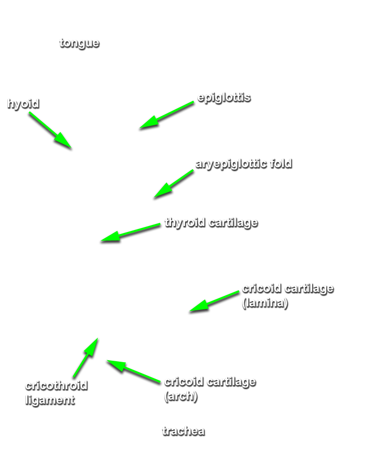
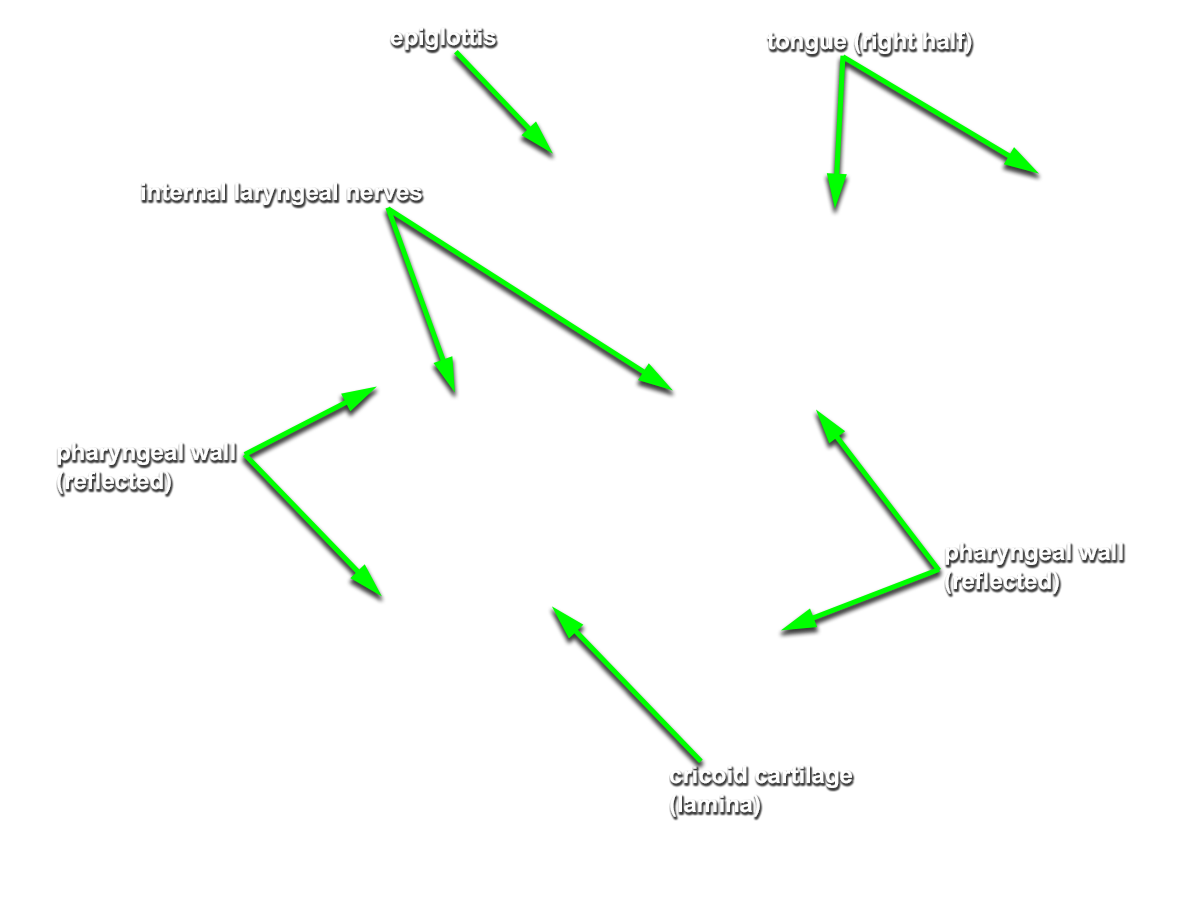
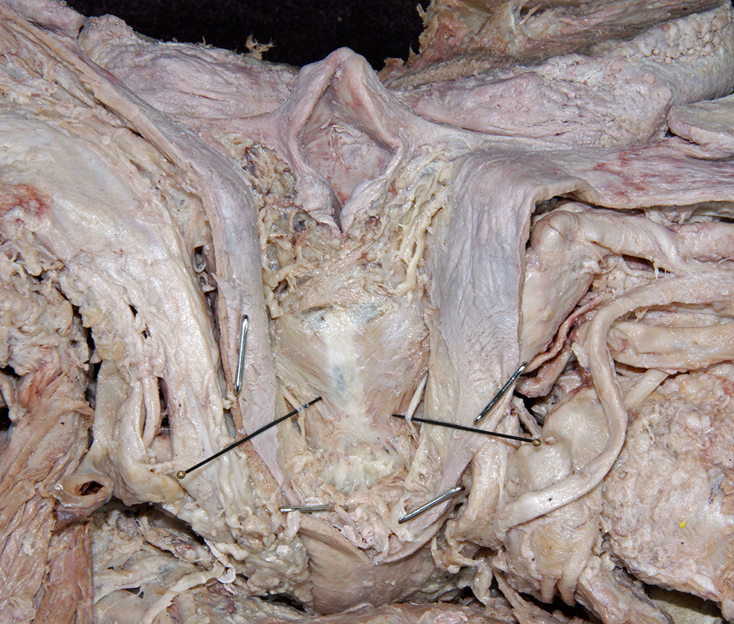
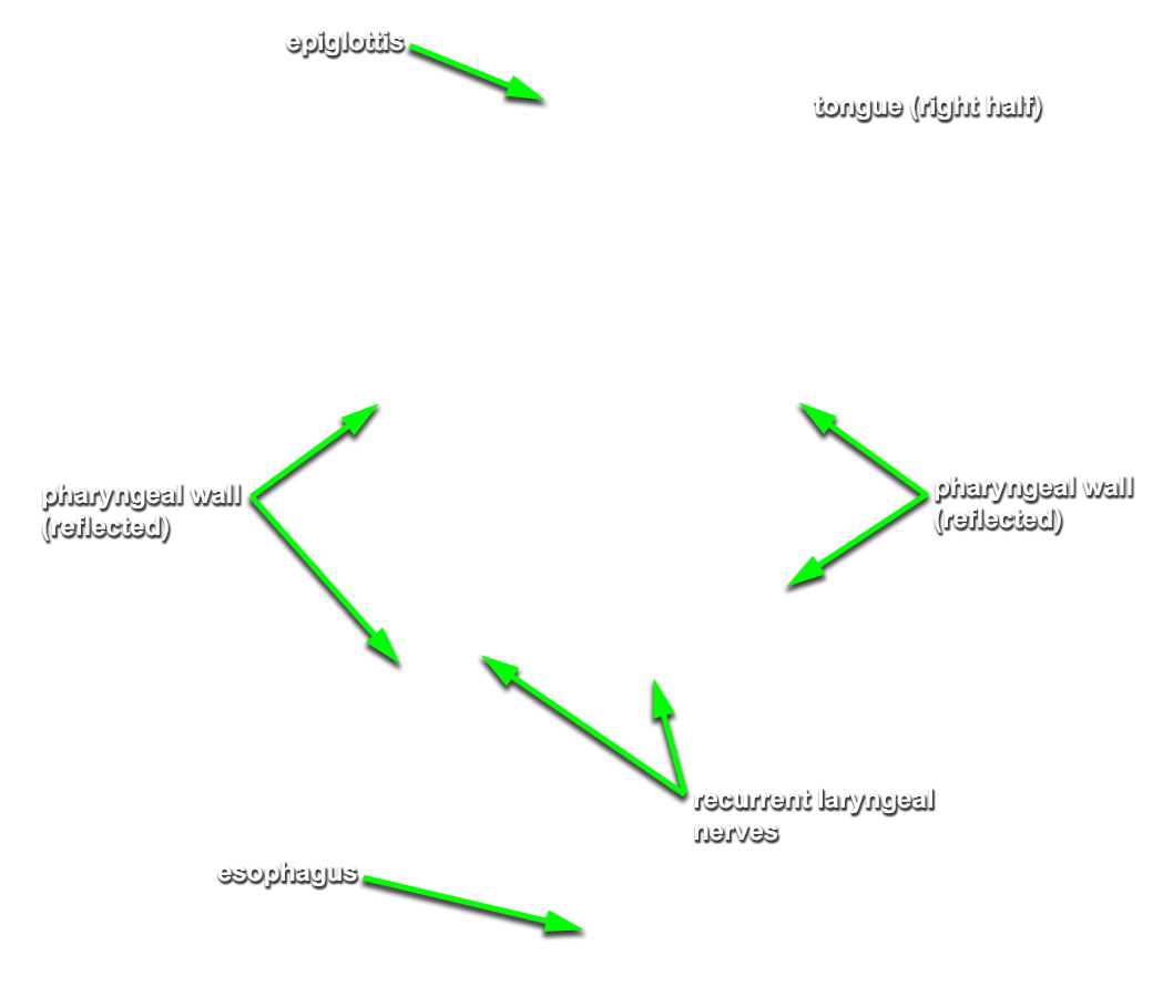
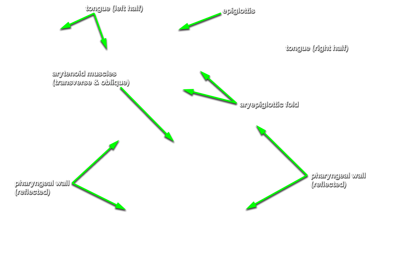
epiglottis,
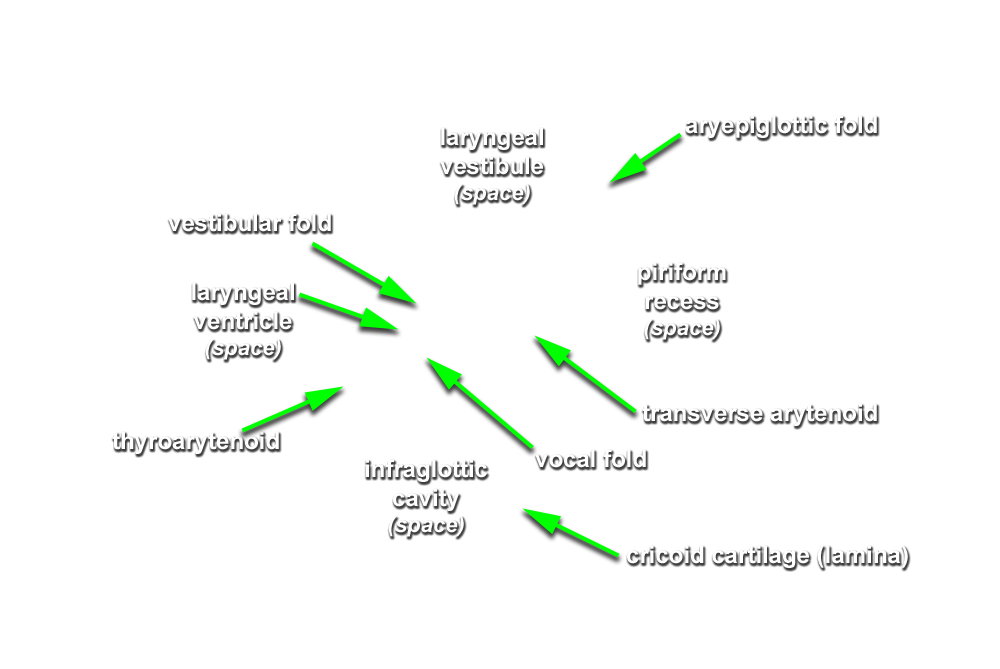
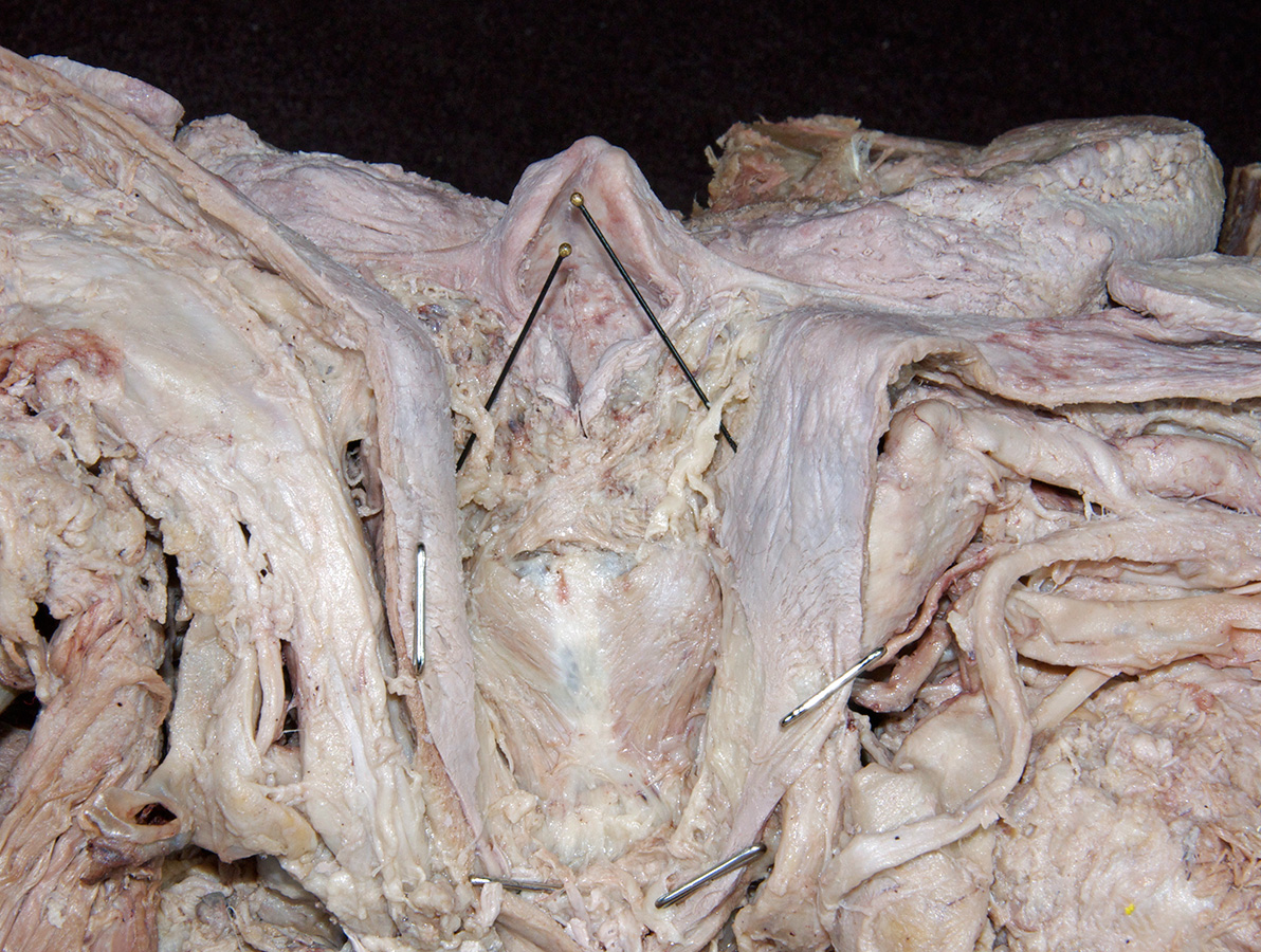
aryepiglottic fold,
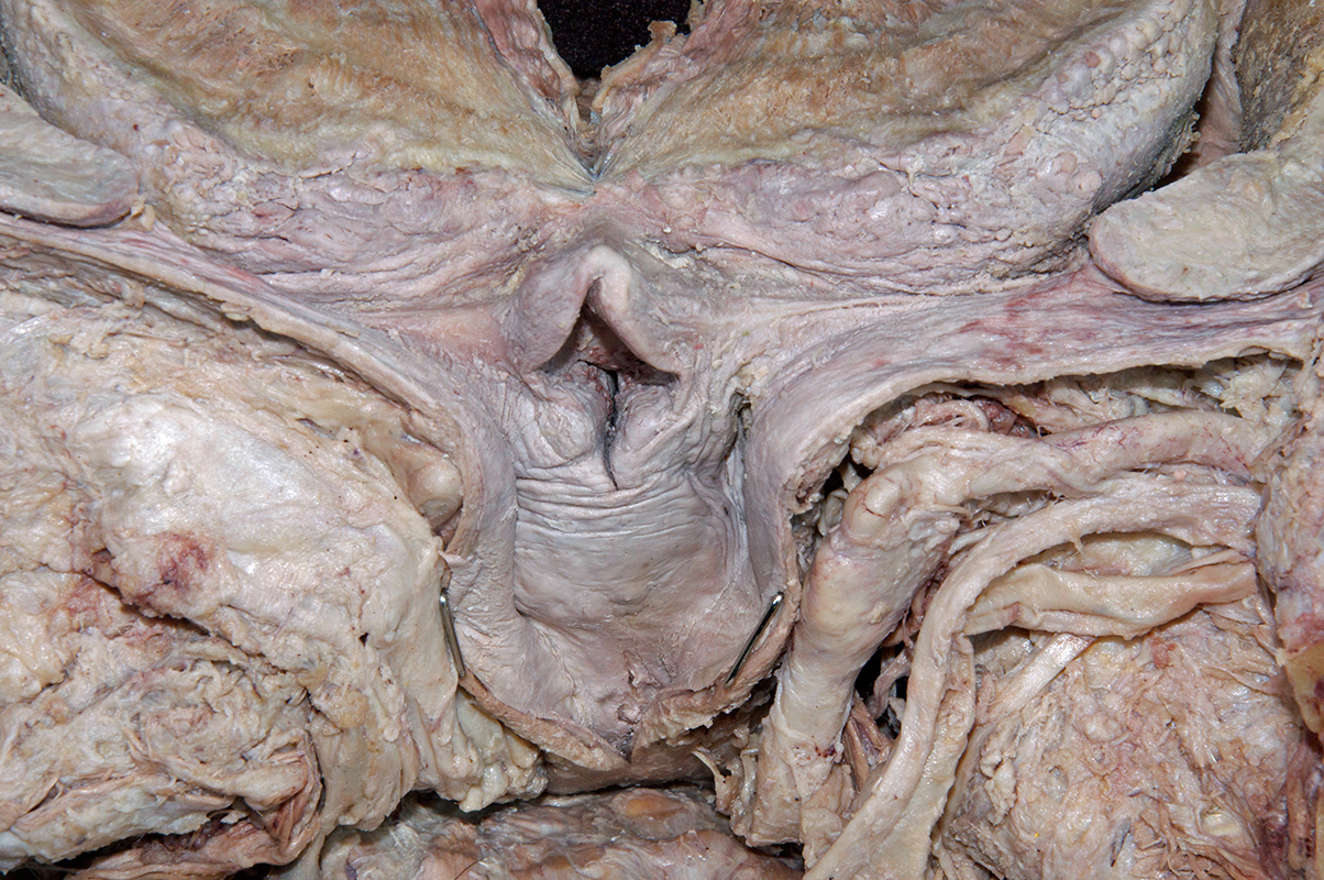
piriform recess,
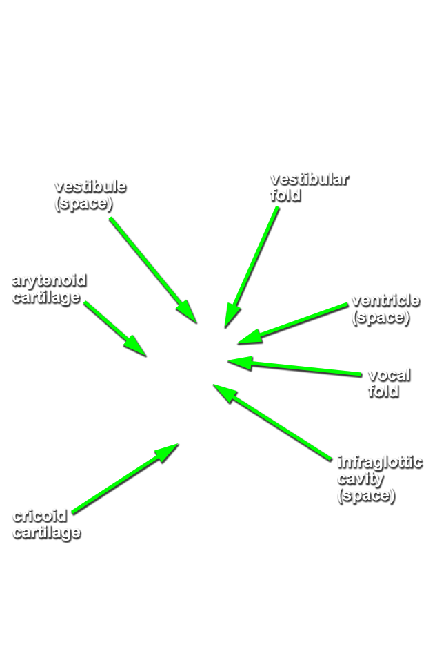
laryngeal vestibule,
vestibular fold,
laryngeal ventricle,
vocal fold and
infraglottic cavity. (G 7.49B;N 58 & 66;Gl 38.2 & 39.20)
Important Relationship
- The vallecula is positioned directly anterior to the epiglottis and posterior to the tongue (root).
- The epiglottis is positioned posterior to the tongue (root).
- The piriform recess is positioned lateral to the laryngeal inlet.
- The vocal ligament is positioned anterior to the arytenoid cartilage.
- The vocal fold is positioned inferior to the vestibular fold.
- Identify the internal laryngeal nerve in the superior aspect of the piriform recess. (G Table 8.10 Posterior View;N 67;Gl 38.34)
- Identify the
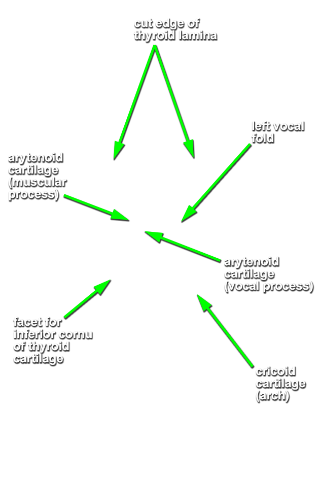
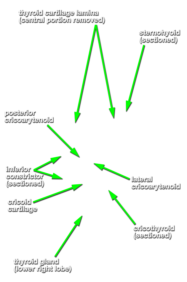
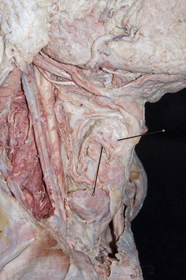
lamina of the cricoid cartilage (G 8.28;N 77;Gl 39.13) and
recurrent laryngeal nerve in the inferior aspect of the piriform recess. (G Table 8.10 Posterior View;N 80)
Important Relationship
- The thyroid cartilage (laminae) are positioned superior to the cricoid cartilage.
- Palpate and identify the
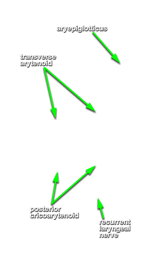
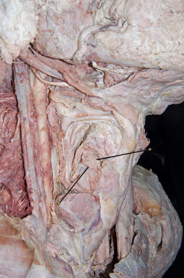
arytenoid cartilage. (G 8.28E;N 77;Gl 39.18B) Identify the
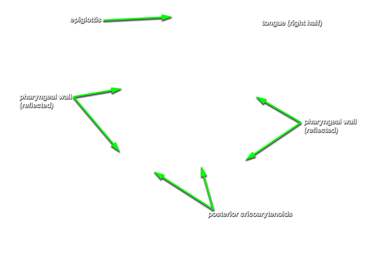
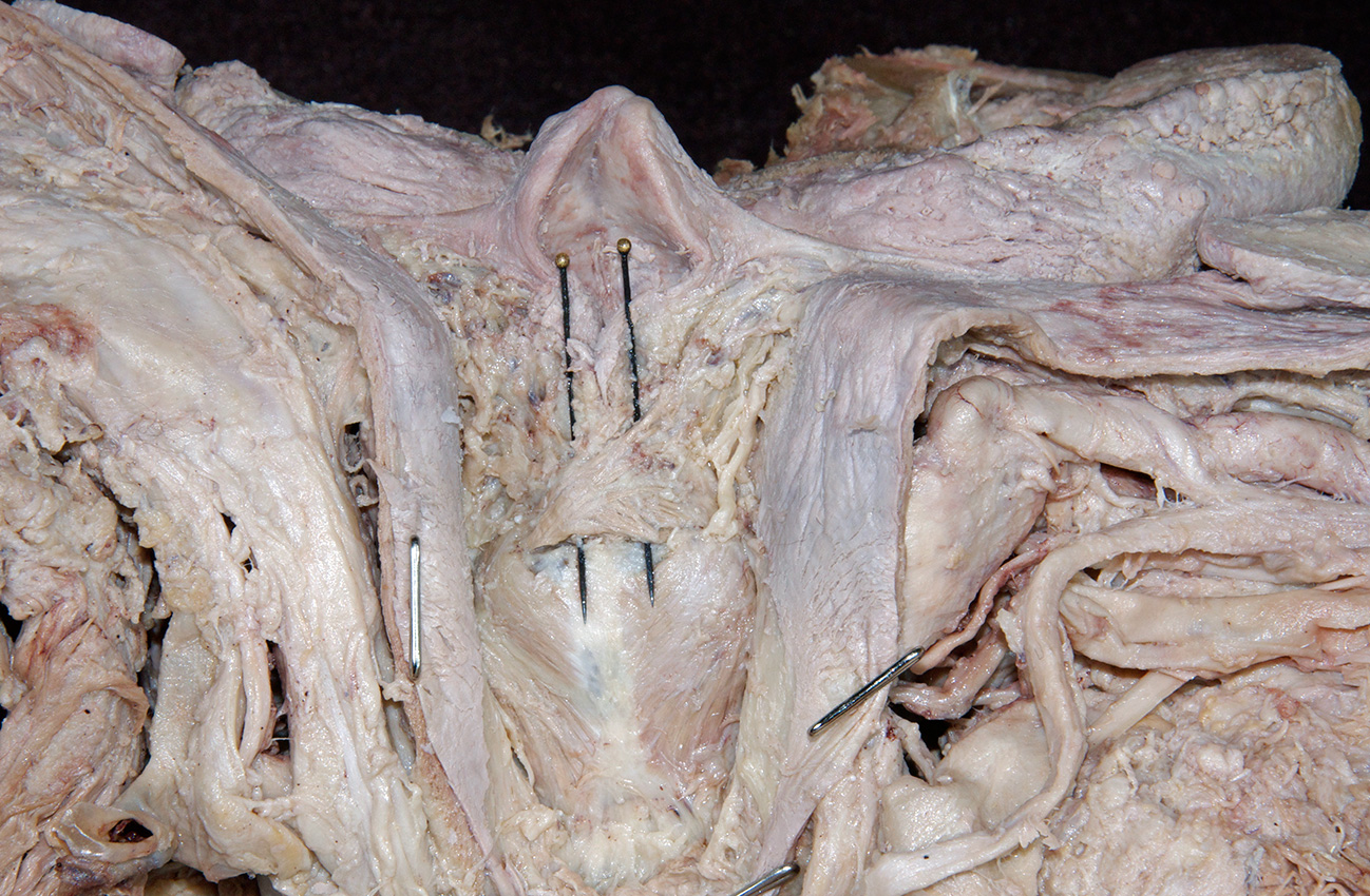
posterior cricoarytenoid and
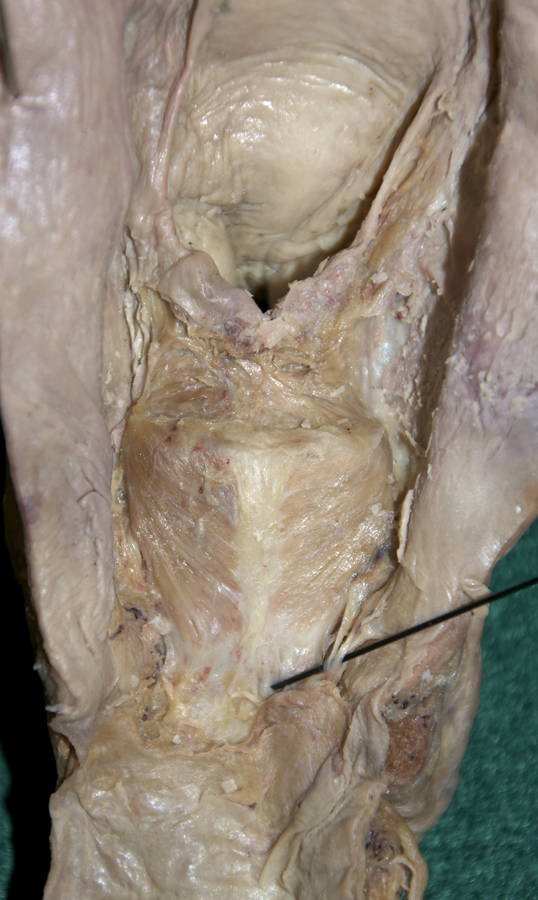
transverse arytenoid muscles. (G Table 8.10 Posterior View;N 78;Gl 38.31C) Attempt to identify the
aryepiglotticus muscle.
Important Relationship
- The arytenoid cartilage is positioned superior to the cricoid (lamina) cartilage.
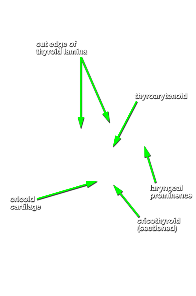
- (OPTIONAL, ON ONE SIDE ONLY - the side where the infrahyoid muscles were cut) Attempt to identify the cricothyroid, lateral cricoarytenoid and thyroarytenoid muscles. (G Table 8.10 Lateral View;N 78;Gl 39.19)