-
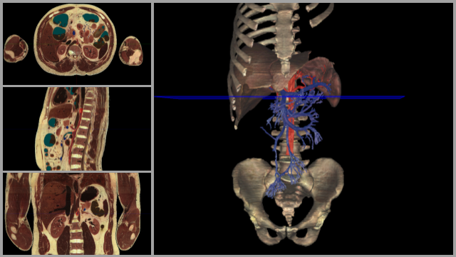
Celiac Trunk & Portal Vein Formation
Rotate the current view and appreciate the extent of the portal and superior mesenteric veins. Can you name all the portions of the GI tract that the superior mesenteric vein travels to? Scroll through the transverse cross sections (using three fingers) and appreciate the branching of the celiac trunk off the abdominal aorta. Notice where you are on the 3D model as you scroll through by noting the position of the blue plane that pans with you as you move your fingers. What are the three branches of the celiac trunk? Can you trace these three branches through the cross section as you scroll through? To gain a better view of the celiac trunk and its branches you can tap the dissect tool at the bottom and remove the veins. Rotate the image again to appreciate the arteries.
-
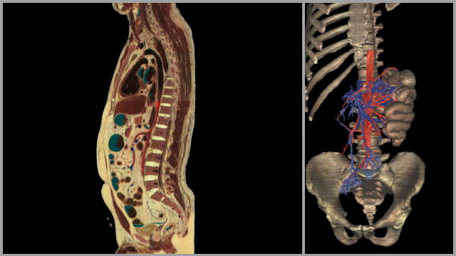
Superior Mesenteric Arteries & Veins
In this view you can see part of the small intestines included (the jejunum). Scroll through the sagittal cross section. Can you see a large vessel traveling inferior to the superior mesenteric artery? Can you identify this vessel? Rotate the 3D view to appreciate where the vessels travel to. If you would like to add more organs to the view select the anatomy tool at the bottom of the screen, under the index tab of the pop up type ascending colon and touch the eye icon next to ascending colon to add it to the view. Do the same with the transverse colon to add it to the 3D view. Rotate the 3D image to appreciate the arterial and venous branches of the superior mesenteric artery and vein.
-
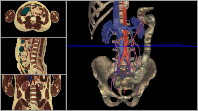
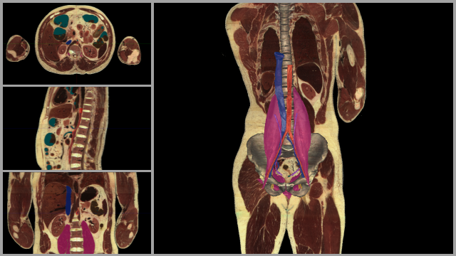
Inferior Mesenteric Arteries & Veins
What portion of the intestine do the inferior mesenteric artery and vein supply? Can you identify where the superior mesenteric artery would branch off the abdominal aorta just superior to the L renal vein? Rotate the 3D image and use the highlight tool to identify the L & R testicular veins. Do you notice a difference in attachment points for these two vessels between the L & R sides? Notice the location where the common iliac arteries and veins branch to form left and right common iliac vessels.
-
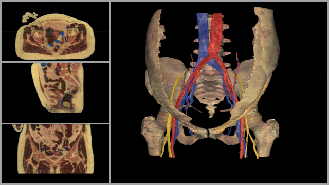
Female Inguinal Region
Double tap on the transverse cross section to enlarge the image. Scroll through the transverse cross section (using 3 fingers) to observe the contents of the inguinal canal. What are the structures within the inguinal canal? Use the dissect tool to look through the layers of abdominal muscles already present in the image.
-
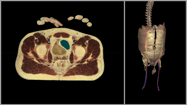
Male Inguinal Region
Scroll superiorly in the transverse cross section to the location where you can see the abdominal muscles. Use the highlight tool to distinguish between the layers of abdominal muscles. Then use the dissect tool to remove the musculature from the 3D image so you can view the structures deeper. Identify the inguinal ligament and highlight it. Find the highlighted inguinal ligament in one of the cross sections. What structures pass through the inguinal canal?
-
Pelvic Floor
Use the highlight and dissect tools, as well as rotation, to help you distinguish between the various muscles of the pelvic floor. Think about the movements that would occur if some of these muscles were to contract. Scroll through the transverse cross section to help you see the relationship between the muscles of the pelvic floor.