-
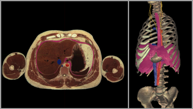
Diaphragm & Structures Passing Through It
Use three fingers to scroll through the transverse cross section. Notice the boundaries of the diaphragm (highlighted in pink) as you move superiorly and inferiorly through the sections. Can you see the location where the esophagus passes through the diaphragm and joins with the stomach? How about the inferior vena cava as it traverses towards the liver? Rotate the 3D view of the anatomy. Can you imagine where the liver resides inferior to the diaphragm? Add the liver by selecting the highlight tool and tap on the liver in the cross section - this will add a highlighted liver to the 3D image. To visualize the structures that pass through the diaphragm better use the dissect tool to remove the diaphragm from view (any any other structures obstructing your view like the liver). What are the various parts of the diaphragm called where each of these structures pass through it?
-

Diaphragm & Nearby Structures - VH Dissector Beta Only
Use three fingers to scroll through the transverse cross section. Notice the boundaries of the diaphragm (highlighted in pink) as you move superiorly and inferiorly through the sections. Can you see the location where the esophagus passes through the diaphragm and joins with the stomach? How about the inferior vena cava as it traverses towards the liver? Rotate the 3D view of the anatomy. Rotate the 3D view so you are looking up under the diaphragm. Locate and highlight (if desired) the liver and spleen. Appreciate their location inferior to the diaphragm. What happens to these structures when you inhale? Use the dissect tool to remove the liver, stomach, and spleen. Can you identify and name the structures passing through the diaphragm and what those passageways through the diaphragm are called?
Previous Lab Review
-
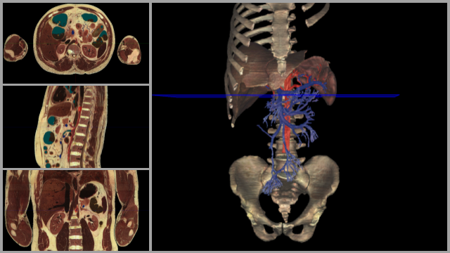
Celiac Trunk & Portal Vein Formation
Rotate the current view and appreciate the extent of the portal and superior mesenteric veins. Can you name all the portions of the GI tract that the superior mesenteric vein travels to? Scroll through the transverse cross sections (using three fingers) and appreciate the branching of the celiac trunk off the abdominal aorta. Notice where you are on the 3D model as you scroll through by noting the position of the blue plane that pans with you as you move your fingers. What are the three branches of the celiac trunk? Can you trace these three branches through the cross section as you scroll through? To gain a better view of the celiac trunk and its branches you can tap the dissect tool at the bottom and remove the veins. Rotate the image again to appreciate the arteries.
-
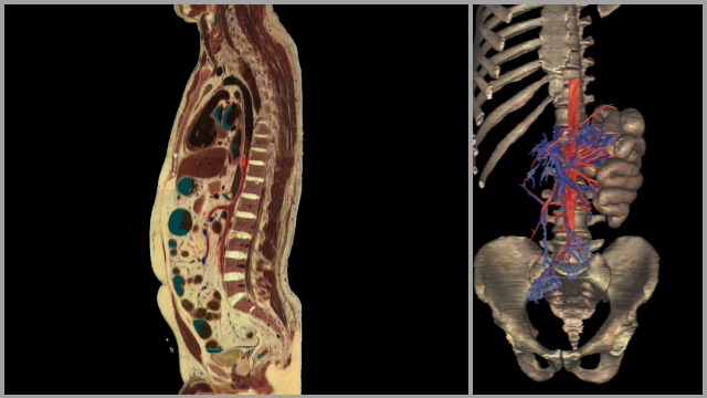
Superior Mesenteric Arteries & Veins
In this view you can see part of the small intestines included (the jejunum). Scroll through the sagittal cross section. Can you see a large vessel traveling inferior to the superior mesenteric artery? Can you identify this vessel? Rotate the 3D view to appreciate where the vessels travel to. If you would like to add more organs to the view select the anatomy tool at the bottom of the screen, under the index tab of the pop up type ascending colon and touch the eye icon next to ascending colon to add it to the view. Do the same with the transverse colon to add it to the 3D view. Rotate the 3D image to appreciate the arterial and venous branches of the superior mesenteric artery and vein.
-
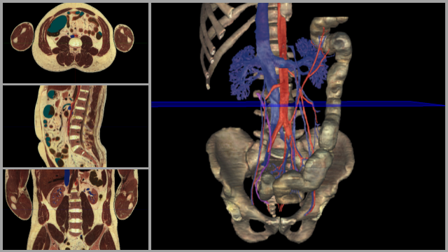
Inferior Mesenteric Arteries & Veins
What portion of the intestine do the inferior mesenteric artery and vein supply? Can you identify where the superior mesenteric artery would branch off the abdominal aorta just superior to the L renal vein? Rotate the 3D image and use the highlight tool to identify the L & R testicular veins. Do you notice a difference in attachment points for these two vessels between the L & R sides? Notice the location where the common iliac arteries and veins branch to form left and right common iliac vessels.