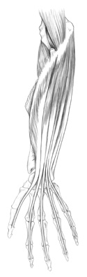
Figure 12-2
Deep Flexors of the anterior forearm.
Deep Flexors of the anterior forearm.
The deep layer of forearm flexors can be identified by their distal attachments and their placement relative to each other. The most superficial and obvious muscle of this group is the flexor digitorum superficialis. Its tendon splits to allow the tendon of the deeper placed flexor digitorum profundus to pass through it. It attaches to the sides of the middle phalanx of digits 2-5.
Now you should easily see the distal attachment of the flexor digitorum profundus on the distal phalanx of digits 2-5.
Deep in the forearm, the muscle just lateral to the profundus arising from the radius and heading to the distal phalanx of the thumb is the flexor pollicis longus.
The deepest muscle of all the flexors in the forearm is the small pronator quadratus. This muscle runs transversely between the distal ends of the radius and the ulna.
Now that you are somewhat familiar with the muscles on this side of the forearm, see if you can find the point slightly distal to the elbow under the pronator teres where the brachial artery splits into the
radial
and
ulnar artery.
Follow the radial artery down to the wrist. It should disappear posterior to the lateral edge of the first metacarpal. If you're lucky, you may be able to see the terminal end of the radial artery, the
deep palmar arch
 .
.
The
ulnar artery
is located on the ulnar or little finger side of the forearm, and should accompany the
ulnar nerve.
At the wrist level, you should be able to see the ulnar artery become the
superficial palmar arch
,
and the ulnar nerve distribute itself to the medial side of the hand and digits.
Lying in the middle of the forearm, midway between superficial and deep layers, find the median nerve. Verify its identity by tracing the distal distribution to the lateral or radial side of the hand and to the thumb.