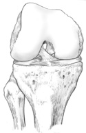
Figure 15-3
Anterior view of the right knee flexed to show the cruciate ligaments and the femoral condyles.
Anterior view of the right knee flexed to show the cruciate ligaments and the femoral condyles.
Working on the medial side of the knee that has been dissected, find the medial or tibial collateral ligaments under the pes anserine. This ligament is somewhat broad and flattened out.
On the lateral side of the knee, find the pencil-like lateral or fibular collateral ligament under the distal end of the biceps femoris as it attaches to the head of the fibula.
On the anterior side of the knee find the patellar ligament and the tibial tuberosity.
Reflect the patella and patellar ligament inferiorly and flex the knee to see the interior of the joint.
Find the anterior cruciate ligament as it arises from the anterior tibial intercondylar eminence and attaches to the medial part of the lateral femoral condyle. (Figures 15-3 and 4)
The posterior cruciate can be seen attaching to the lateral portion of the medial femoral condyle. You may be able to see the posterior cruciate from a posterior approach through the popliteal fossa.
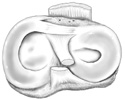
Now observe the two cartilaginous menisci or circular crescent shaped rings of cartilage. Can you see how they are thicker at their edges and incomplete in the center, so they resemble a saucer without any middle? Now you can appreciate how these cartilaginous rings act as shock absorbers and stabilize the knee by deepening the tibial table-top.
Note how the C-shaped medial meniscus is firmly attached to the tibial condyle and to the medial collateral ligament of the knee. (Figure 15-4)
Notice how the O-shaped lateral meniscus has no attachments to the lateral collateral ligament and is much more mobile than the medial meniscus.