Figure 16-4
Arcuate Artery.
Arcuate Artery.
Looking at the dorsal surface of the foot and deep to the tendons of the extensor digitorum longus and peroneus tertius, find the small single muscle on the dorsum of the foot, the extensor digitorum brevis. Some anatomists will single out the medial most fibers and tendon of this muscle and call it the extensor hallucis brevis.
Follow the
deep peroneal nerve
onto the dorsal surface of the foot. It usually follows the same path as the anterior tibial artery. The anterior tibial artery, at the level of the talocrural joint, gives off malleolar branches and becomes the
dorsal pedis artery.
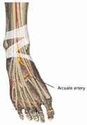
You may be able to see the dorsal pedis artery swing laterally to become the
arcuate artery
 or dorsal arch. The arcuate artery gives rise to the dorsal digital branches.