Previous
|
Index
|
Next
2: Thoracic Wall and Contents, Heart and Lungs
Introduction
-
Overview
-
Identification
-
Summary
-
Imaging
-
Clinical Case Study
X-Ray Imaging of the Thorax
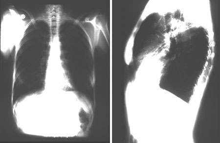
Figure 2-20
X-rays of the thorax. The left image is a frontal exposure and the right image is a lateral exposure.
Identify 5 significant structures on these X-rays.