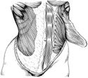
Figure 3-10
Oblique muscles of the lower thorax and abdomen. External oblique elevated on left side to expose internal oblique.
Oblique muscles of the lower thorax and abdomen. External oblique elevated on left side to expose internal oblique.
Begin by identifying the aponeurosis of the external intercostal muscles of the rib cage and the external abdominal oblique muscles. Observe the external abdominal oblique muscle running from the lower eight ribs medially and inferiorly, just like the external intercostals above.
Directly beneath this muscle find the internal abdominal oblique muscle, with its muscle fibers running inferiorly and lateral at right angles to the external oblique. (Figure 3-10)
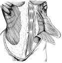
Continue your journey inward through the abdominal wall by finding a third deeper layer composed of the transversus abdominis muscle. This muscle should be running nearly in a transverse plane. Deep to the transversus see if you can single out the deepest layer of the wall the transversalis fascia.
Now examine the extent of the right and left sections of the rectus abdominis. Note the transversely oriented tendinous inscriptions on the rectus abdominis. These connective tissue bands not only segment the muscle,(could these be remnants of ribs?); they also attach to the rectus sheath insuring a solid interaction between all of the muscles of the abdominal wall. (Figure 3-11)
At the end of this exercise when the abdominal viscera have been removed, the muscles of the posterior abdominal wall will be explored.