 on the inferior surface of the diaphragm?
Begin at the most superior end of the abdominal cavity. Find and examine the extent of the
diaphragm.
Can you see any
parietal peritoneum
on the inferior surface of the diaphragm?
Find the liver in the right upper abdominal quadrant directly inferior to the diaphragm. It is easy to identify because it is the largest gland in the body and fills almost all of the upper right quadrant of the abdomen. It is not uncommon for the liver to fill at least half of the upper left quadrant. The inferior surface of the right lobe of the liver contains the green gallbladder lodged in a fossa. Its surface may be attached to the liver by connective tissue and vessels.
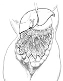
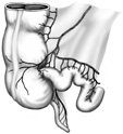
Another prominent structure of the abdomen is the
greater omentum
.
This double layer of peritoneum arises from the greater curvature of the
stomach
(the stomach may be dilated and conspicuous or contracted and less evident). It is draped inferiorly over the most of the intestines allowing only a part of the descending and sigmoid colons to show. The greater omentum is a filmy apron containing a large amount of fat distributed between the blood and lymph vessels and and lymph nodes. (Figure 3-13)
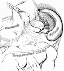
Find the esophagus, a muscular tube passing through the superior and posterior mediastinum and diaphragm to enter the stomach at its cardiac orifice.
Identify the greater and lesser curvatures of the stomach as you explore it. At the inferior or distal end of the stomach feel the "gatekeeper" of the stomach, the pyloric sphincter. The pyloris is the opening of the stomach into the duodenum. It controls the flow of stomach contents into the duodenum. (Figure 3-14)
Look for the spleen posterior to the stomach and inferior to the diaphragm in the upper left quadrant under the ribs; note the large blood vessels entering and leaving the organ. (Figure 3-15)
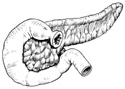
Examine the duodenum. Note its C-shape and how it is molded to the head of the pancreas. The pancreas is somewhat diffuse and hard to recognize. By remembering that it is held in the "arms" of the duodenum, you will always be able to find it.
The duodenum continues for only about a foot before the transition into the jejunum. The section identified as the duodenal-jejunal junction takes a sharp bend inferiorly near the inferior border of the pancreas. (Figure 3-16)
The distal end of the small intestine, the ileum, is most easily identified as it enters the cecum, at the ileocecal junction. This junction is in the inferior right quadrant of the abdomen. (Figure 3-17)
Now is a good time to find and explore the
root of the mesentery
.
The mesentery stretches diagonally across the posterior wall from the duodenal-jejunal junction along the jejunum and ileum to the ileocecal junction. It is only 6 or 7 inches long. This double layer of peritoneum sandwiches the superior mesenteric artery and vein and suspends the small intestines from the posterior abdominal wall. (Figure 3-18)
Try putting one hand on either side of the mesentery and drawing your fingers anteriorly. This will effectively untwist some of the intestinal convolutions to make the duodenal and cecal ends more obvious.
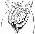
Turn your attention to the large intestine. Remember, the cecum is the very beginning of the large intestine and is a blind pouch or sac. See if you can find an appendix attached to the cecum. Usually it does so on the posterior side of the cecum.
Follow the ascending colon superiorly to a point where it takes a right angle turn.
The transverse colon begins here and continues until it takes another right angled turn near the border of the spleen.
The transverse colon now becomes the descending colon. It runs inferior to the pelvic brim taking an S-shaped turn toward the midline. This S-shaped portion is called the sigmoid colon. It continues to near the midline, takes a sharp right angled turn inferiorly and becomes the rectum. (Figure 3-18)
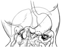
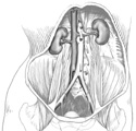
Your next step is to explore the superior aspect of the posterior abdominal wall and locate the kidneys. Notice how the kidneys lie right against the posterior abdominal wall just barely under the last ribs. Also notice how the left kidney lies superior to the right kidney.
At the root or hilus of the kidney notice there are three structures: an anterior renal vein, a more posterior renal artery, and a ureter. Follow the ureter inferiorly and observe it cross the pelvic brim and enter the urinary bladder. (Figure 3-19)