Abdominal Wall
The organs of the abdominal cavity are protected anteriorly and laterally by the multi-layered abdominal wall consisting of skin, fascia, muscle and serous membrane.
The abdominal muscle arrangements are surprisingly similar to the muscle arrangements of the thorax and back. The external and internal abdominal oblique muscles are oriented in exactly the same pattern as the external and internal intercostal muscles, however there are no ribs. The transversus abdominis can be likened to the transversus thoracis. The longitudinal rectus abdominis can be an anterior erector spinae and vertebral column.
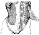
The abdominal obliques and transverse abdominis , called "flat" muscles converge, anteriorly, in the connective tissue "aponeurosis" on and around the rectus abdominis forming the rectus sheath. The muscles are bilateral with each side converging on the midline to form a tough white connective tissue band called the linea alba. The linea alba is important clinically. Surgical incisions can be performed through it without significant damage to nerves or blood vessels. (Figure 3-2).
The deepest layer of the abdominal wall is the deep fascia of the transversus abdominis or the transversalis fascia. This fascial layer lines the inside of the abdominal wall.
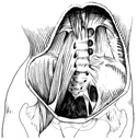
The posterior abdominal wall is made up of several muscles that maintain posture, flex the hips and help keep them level while walking. These muscles include: the psoas, which runs from the inside of the lumbar vertebra to the femur, the quadratus lumborum, extending from the hips to the lower ribs and vertebral column and the posterior portions of the transversus abdominis. (Figure 3-3)
The ventral rami of spinal nerves T6-T12 and the L1 spinal nerve supply the abdominal wall muscle and skin with motor and sensory nerve fibers.
The blood supply to the abdominal wall really mimics that of the thoracic wall. Arterial blood is delivered to the anterior abdominal wall by:
- The terminal end of the internal thoracic artery, the superior epigastric.
- The ascending inferior epigastric artery from the external iliac artery in the pelvic cavity.
The posterior abdominal wall receives arterial blood from four pairs of lumbar arteries arising from the descending aorta much like the posterior intercostal arteries of the thorax.
Venous return is also similar to that of the thoracic wall; there is a separate posterior set of veins, part of the azygous system, and epigastric veins that empty the anterior abdominal wall.
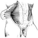
In each of the two sexes, there is an anterior abdominal wall swelling containing structures related to the urogenital system.
In the male, the testes descend in the abdomen and are drawn outside the abdominal wall by their embryonic connection, the gubernaculum, to the outpouching of the anterior abdominal wall, the scrotum. The testes, however, still retain their abdominal connections by way of the spermatic cord. This cord can be thought of as the "life line" of the testes, as it contains blood vessels and the duct of the testes, the vas deferens.
The spermatic cord is composed of an inner core containing the spermatic arteries, veins and lymphatic vessels; with testicular nerves and the ductus deferens. A laminated fascia called the internal spermatic fascia is continuous with the transversalis fascia at the deep inguinal ring. It encases the inner core. The inner core is covered by the cremaster layer consisting of scattered bundles of the cremaster muscle. The cremaster muscle is continuous with the internal oblique muscle and its fascia in the abdominal wall. The inner core and the cremaster layer are encased in a thin membrane prolonged distalward over the cord and testis. It is continuous, at the superficial inguinal ring, with the deep fascia covering the external oblique muscle of the abdomen. (Figure 3-4)
The descent of the testes can be visualized by imagining the testes pushing through the anterior abdominal wall during development. They push all layers of the wall ahead of them until they become completely invested in the scrotum, an external pouch suspended from the abdominal wall.
In the female, the ovaries do not descend so far. They remain within the abdominal cavity. However, they too have a connection to the anterior abdominal wall outpouching. The round ligament of the uterus extends from the superior part of the lateral border of the uterus to the pelvic wall. There it penetrates the abdominal wall via the deep inguinal ring. It passes through the inguinal canal and its fibers spread out to attach to the labia majora.
In both sexes, holes exist in the lower quadrants of the anterior abdominal wall where either the spermatic cord or the round ligament of the uterus pass. They are known as inguinal canals because they pass through all the layers of the abdominal wall forming short canals. The canals can be problem sites. Inguinal hernias, the protrusion of abdominal contents through the canal, are among the most common.
In the male, the spermatic cord and its relationship to the inguinal canal are clinically significant in cases of infertility and inguinal hernias.
In the female, inguinal hernias are less frequent because the inguinal canal contains only the round ligament of the uterus and is smaller in size.