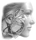
Figure 6-1
Facial nerve VII and associated structures.
Facial nerve VII and associated structures.
As you work in this area note how the muscles of expression are nearly the same color as the rest of the surrounding fatty and connective tissue. These muscles are very thin, attaching to the skin of the face from the superficial fascia or the facial bones.
They all receive motor innervation from cranial nerve VII, the facial nerve. The motor portion of cranial nerve VII exits the skull via the stylomastoid foramen, runs through the parotid salivary gland and then divides into 5 or so branches that supply the muscles of expression (Figure 6-1).
The parotid gland (x2) located just anterior and slightly inferior to the external ear, is a multilobar gland; it has many individual lobules and looks a little like a sponge. The parotid duct carries the secretion of this gland (saliva) to the mouth, emptying behind the second upper molar. Injury or disease of the parotid gland can be very debilitating as this could affect cranial nerve VII and cause the loss of expressive muscles. Because of the proximity of cranial nerve VII to the external and middle ear, problems in this region can also have an affect on cranial nerve VII. See case history in this exercise.
In the superior portion of the neck, you will be looking for the common carotid artery and its branches. This artery runs with cranial nerve X, the vagus nerve, and the internal jugular vein and all three are surrounded by a connective tissue "sheath," the carotid sheath. Deep to, or sometimes included in, this neurovascular bundle you may be able to see the sympathetic trunk. The common carotid, at the level of our adam's apple or so (C4), splits into an internal carotid, which supplies the brain, and an external carotid, which supplies almost everything else in the head. In this exercise we will look at the external carotid as it gives off branches to the neck, tongue, face, teeth, nasal cavities and scalp.
Remember, the muscles of mastication are exclusively involved with movement of the temporomandibular joint. They facilitate chewing food, swallowing, breathing and talking. Also remember these muscles will have different functions if they act bilaterally or unilaterally.
You probably will not see their motor supply from the mandibular division (V3) of the trigeminal, cranial nerve V. You will see the sensory branches of the cranial nerve V as they leave the skull on their journey to the tongue, mouth, teeth and gums and the skin of the face and head.
Keep in mind the trigeminal nerve has three divisions: