Previous
|
Index
|
Next
7: The Brain, Cerebral Arterial Circle, Dura Mater, Venous Sinuses and Cranial Fossa
Introduction
-
Overview
-
Identification
-
Summary
-
Imaging
-
Clinical Case Study
Imaging
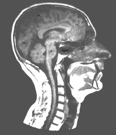
Figure 7-14
Midsagittal MRI image of the head. (PL) parietal lobe, (FL) frontal lobe, (OL) olfactory lobe, (C) cerebellum, (P) pons, (SphS) sphenoidal sinus.