Definition of Portal Hypertension
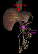
Injuries to the
liver
lead to an obstruction in portal flow and result in elevation of the portal venous pressures. In
addition, a decrease in
portal flow
leads to the release of endogenous vasodilators, resulting in an increase in
hepatic arterial flow
into the liver. This can lead to a further increase in portal venous pressure.
Normally the pressure difference between the portal circulation and the systemic circulation is
5-10 mm Hg. In some literature, portal hypertension exists when the portal-systemic venous gradient
is greater than 12 mm Hg, with the portal pressure estimated by the hepatic vein wedge pressure.
Other reports define portal hypertension as more than 5 mm Hg above the systemic pressures.
Causes of Portal Hypertension
The most common cause of portal hypertension is cirrhosis of the liver, with a relative obstruction
to flow. Other causes are increased flow in the portal circulation as seen with arterio-venous
fistulae, obstruction to liver drainage (restrictive pericarditis, hepatic vein thrombosis) or
obstruction below the liver (portal vein thrombosis, splenic vein thrombosis).
Anatomic Results of Portal Hypertension
Portal hypertension leads to the development of and blood flow through collateral vessels.
Clinically, the
most important of these are related to the gastric veins and vessels surrounding the
esophagus,
which flow to portal systemic shunts in the proximal esophagus. The
short gastric veins
arise off of the
splenic vein.
A posterior view can better demonstrate the relationships when the
stomach
is superimposed. Finally, investigate the
stomach and esophagus
from a lateral view to better appreciate the splenic and gastric veins.
Clinical Manifestations
The spontaneous shunts and the resultant enlargement of vascular channels in the stomach and
esophagus (esophagogastric varicies) are the most important clinical manifestation of portal
hypertension. The resultant varicies frequently rupture leading to life-threatening hemorrhage.
Rectal shunts with resultant rectal varicies can also develop. Below are video clips showing
normal anatomy, as well as images from patients with varicies.
Esophageal Varicies.
Gastric Varicies.
Endoscopy of Normal Esophagus.
There are still other manifestations of portal hypertension. Fetal vascular structures
in the
falciform ligament
(superior border shown), such as the residual
umbilical vein,
can dilate with resultant abdominal wall varicosities
(anterior view),
as well as splenic-renal vein anastomoses
that can lead to retroperitoneal varicies.