As discussed in Lesson 1, portal hypertension is caused when there is a relative obstruction to
portal venous flow,
generally at the level of the liver,
as blood moves into the systemic circulation through the
hepatic veins and
inferior vena cava.
Portal hypertension leads to clinical problems, which include gastrointestinal bleeding and ascites.
A variety of surgical methods have been developed to decompress the
portal circulation
directly into the
systemic circulation,
generally following a gastrointestinal hemorrhage of either
esophageal
or
gastric varicies.
These decompressive measures are generally very effective for preventing rebleeding.
However, since blood is diverted away from the liver,
encephalopathy tends to develop as the bypassed liver fails to prevent toxins from reaching the systemic
circulation.
Shunts can be total, partial or selective.
They can be created during incisional surgery, or placed radiologically.
Portosystemic Shunts decompress all parts of the portal circulation and deprive the
liver of portal blood flow. Partial Portosystemic Shunts limit the shunt flow (lumen of shunt less than 8 mm) and preserve
some portal flow into the liver.
Selective Portosystemic Shunts, such as the Distal Splenorenal Shunt, decompress only a
part of the portal circulation.
A radiologically placed total (and sometimes partial) shunt known as the Transjugular Intrahepatic
Portal Systemic Shunt (TIPSS)
has become popular over the past 10 years and is probably the most common portal shunting procedure
in use at the time of this writing.
In this lesson, the following shunts will be illustrated:
- End-to-Side Portacaval Shunt
- Side-to-Side Portacaval Shunt
- Central Splenorenal Shunt
- Partial Portal Systemic Shunt
The next lesson (Lesson 3) will cover:
- Distal Splenorenal Shunt
- Transjugular Intrahepatic Portal Systemic Shunt
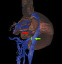
End-to-Side Portacaval Shunt
This total shunt involves division of the
portal vein at or near the hilum
of the liver. The proximal portal vein is ligated, and the distal portal vein is sewn into the
inferior vena cava.
The liver can not be decompressed through the portal vein, and both ascites and encephalopathy can worsen with this type of surgery.
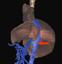
Side-to-Side Portacaval Shunt
This shunt creates a channel between the
portal vein and the
inferior vena cava. The
liver can decompress through the
portal system, so less ascites are
anticipated, but otherwise results are similar to those obtained with end-to-side anastomoses.
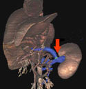
Central Splenorenal Shunt
This shunt theoretically could be a partial shunt, and maintain some portal flow to the liver.
The
spleen, shown from the posterior aspect, is removed and the
splenic vein anastomosed to the
renal vein.
Initially, the shunt may be less than 8 mm in cross section, making it a partial shunt.
But over time, the shunt either enlarges into a total shunt, or thromboses.
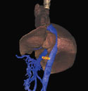
Partial Portal Systemic Shunt
Partial Portal Systemic Shunts employ grafts to prevent enlargement of the shunt.
These grafts are generally 8 mm in diameter and go between the
portal vein and the
inferior vena cava.
The graft keeps the lumen at a small enough size to provide some blood flow through the liver,
but is large enough to deter thrombosis.