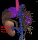
Distal Splenorenal Shunt
Selective Distal Splenorenal Shunt is probably the most widely used surgical portal decompressive procedure.
It is primarily used for
esophagogastro-variceal hemorrhage,
with control of bleeding achieved in the majority of patients.
In this procedure, the
splenic vein
is ligated off of the
portal vein/superior mesenteric vein
and reattached to the
renal vein,
diverting the splenic venous effluent directly into the systemic circulation.
Some of the veins which normally drain the stomach into the portal system near the portal confluence,
including the
left gastric vein, are tied off.
The rest of the
gastric venous system,
(and the attached esophageal varicies), are selectively decompressed through the splenic vein
into the systemic circulation via the
renal vein
and
vena cava.
However, the remainder of the
portal venous circulation, is not decompressed.
This preserves some portal flow to the
liver.
The figure above shows a Distal Splenorenal Shunt.
The
inferior mesenteric vein
should not be ligated when the left gastric vein is tied off,
but should continue to drain into the splenic vein (as also shown in the figure).
Since some portal flow to the liver is maintained, encephalopathy is theoretically lessened with this surgical procedure.
However, the results of comparative trials assessing the rate of encephalopathy and other adverse outcomes between this shunt and portocaval shunts have given mixed results.
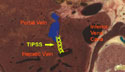
Transjugular Intrahepatic Portal Systemic Shunt (TIPSS)
Transjugular Intrahepatic Portal Systemic Shunt,
TIPSS (generally from the right hepatic to right portal vein) have the advantage of being placed without the need for an abdominal incision.
TIPSS have a high rate of technical success with good short term results.
Long term TIPSS failure is common due to stenosis and thrombosis of the shunt.
These shunts may not be as good as Selective Splenorenal Shunting in the long run,
but are safer to do initially.
TIPSS are generally considered complete shunts (anastomosis greater than 8 mm in diameter),
rather than partial shunts.
Encephalopathy can occur or worsen after a TIPSS.
In contrast to the Selective Distal Splenorenal Shunt and the Portal Systemic Shunts,
TIPSS is preformed by interventional radiologists rather than surgeons.
The right jugular vein
is cannulated and a catheter is passed through the
subclavian vein,
brachiocephalic vein,
the superior vena cava,
the right atrium,
the inferior vena cava
and out into the right hepatic vein.
A wire is passed through the parenchyma of the right lobe of the liver
into the right portal system
and a stent is passed over the wire connecting the hepatic vein to the portal vein.
The stent can be dilated to bring the portal pressures to within 10-12 mm Hg of the systemic venous pressures.
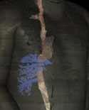
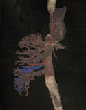
Figure 2 and 3 show the manipulations of a theoretical wire into the right hepatic vein
(Figure 2) and into the right portal vein (Figure 3).
The figure below shows cross sectional anatomy of a TIPSS going from the right hepatic vein into the right portal vein.