The esophagus is a tubular structure situated in the thorax and neck. The caudal part of the esophagus goes through the diaphragm into the stomach. The cephalad portion of the esophagus begins at the upper esophageal sphincter near the cricoaretenoid muscles and has a short cervical section prior to entering the thorax between the superior lobes of the lungs.
For cancer staging purposes, the esophagus is divided into four segments: the cervical esophagus (above the thoracic inlet), the upper thoracic esophagus (from the thoracic inlet to the tracheal bifurcation), the mid thoracic esophagus (from the tracheal bifurcation to a point approximately 8 cm distally) and the lower thoracic esophagus (the remaining esophagus to the gastroesophageal junction). The portion of the esophagus involved becomes important during nodal staging (see below).
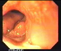
There are two main histologic types of cancer which arise in the esophagus. The first is squamous cell cancer, which appears to be related to tobacco, alcohol and dietary factors. The other is adenocarcinoma, which appears to be related to injury associated with chronic reflux of gastroduodenal contents. An example of a distal esophageal adenocarcinoma is shown in the video.
Therapy of esophageal cancer is determined by the stage of the cancer. Surgery is performed for early stage disease, surgery plus additional therapy for more advanced local disease, and palliative treatment for incurable metastatic disease. Staging determines a T stage (tumor), an N stage (Lymph nodes) and an M stage (metastasis). Preoperative staging is referred to as clinical staging. Procedures used in clinical staging include physical examination, endoscopy, endoscopic ultrasonography, imaging studies using barium, and CT scanning. Pathological staging is done at the time of surgery and is noted with a small case "p".
Although TNM staging of the esophagus generally follows rules used for other GI malignancies, esophageal cancer staging does have some unique features. Tumor (T staging) is somewhat different in esophageal cancer since there is no serosa surrounding the esophagus. The following are the TNM-stages .
T1: Tumor involves the lamina propria or submucosaT2: Tumor involves the muscularis propria
T3: Tumor involves the adventitia
T4: Tumor involves adjacent structures
The adjacent structures which are commonly involved with T4 lesions include the aorta, pleura/lung and pericardium/heart.