Bones of the Shoulder
We will begin by building a shoulder starting with the bony components of the right shoulder. The cross section is at the level of Vertebra T2. (NOTE: You are looking from inferior to superior at the transverse cross sections, so structures on the body's right appear on the LEFT side of the transverse cross section. This is standard radiological presentation.)
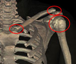
- Moving to the posterior view, place the Scapula on the ribs.
- Rotate the torso laterally and add the Humerus.
-
Move anterior to add the
Clavicle and the
Sternum.
- The Clavicle and the Scapula form the 'shoulder girdle' to attach the arm to the trunk.
The Shoulder Girdle
-
Anterior:
-
Posterior:
-
Scapulothoracic Articulation
- Attaches the shoulder girdle posteriorly to the trunk.
- This attachment is formed by the Subscapularis muscle and the thoracic cage.
The clavicle serves as a "strut" to stabilize the orientation of the shoulder at the Acromial end of the Scapula. The Acromioclavicular (A-C) Joint is supported by the strong
Coracoclavicular Ligament and the
Coracoacromial Ligament. The
Subacromial Bursa is inferior to the A-C joint. The Head of the Humerus fits into the Glenoid Fossa and its extension, the
Glenoid Labrum, to form the Glenohumeral or true 'shoulder joint' that is enclosed by the
Shoulder Joint Capsule.
Muscles of the Shoulder
Now we will build the muscular stabilizers of the
left shoulder. As we continue, use the transverse sections to identify the muscle origins and insertions in order to understand the direction of forces generated when the muscle contracts.
Six Scapular Stabilizers
The Scapulothoracic Articulation is stabilized by 6 extrinsic shoulder muscles that originate from the axial skeleton and insert on the Scapula.
-
Deep Posterior Muscles: (click here to see a labeled image of the deep muscles)
-
Levator Scapulae
- Descends from the Transverse Processes of the Cervical Vertebrae to insert on the Superior Angle of the Scapula.
- Functions as an accessory scapular elevator.
-
Rhomboid Minor and Rhomboid Major
- Descend from the Cervical Spine and upper thoracic spinal ligaments to insert on the Medial Border of the Scapula.
- Function as scapular adductors.
-
Superficial Posterior Muscle:
-
Trapezius
- Upper Trapezius originates from the Cervical Spine and inserts on the Clavicle and the Acromion.
- Middle Trapezius originates from the lower Cervical and upper Thoracic Spine and inserts on the Scapular Spine.
- Lower Trapezius originates from the lower Thoracic Spine and inserts on the inferior portion of the Scapular Spine.
- Functions as a major stabilizer.
-
Lateral Muscle:
-
Serratus Anterior
- Arises from the Ribs and inserts on the Medial Border of the Scapula.
- Functions synergistically as a Scapular protractor and rotator.
-
Anterior Muscle:
-
Pectoralis Minor
- Originates from the anterior of ribs 2-5 to insert on the Coracoid Process of the Scapula.
- Functions with the Serratus Anterior to draw the Scapula anterolateral around the chest wall. It also assists in rotation of the Scapula (with the Levator Scapulae and the Rhomboids) to depress the shoulder.
The Rotator Cuff
Four musculotendinous units form the 'rotator cuff' which provides the dynamic muscular tension to pull the Humeral Head into the shallow Glenoid Fossa for joint security during movement. The "SITS" muscles that form the Rotator Cuff are the Supraspinatus, Infraspinatus, Teres Minor and Subscapularis.
-
Supraspinatus (note cross section)
- Originates from the Supraspinous Fossa of the Scapula, extends under the Acromion, and inserts on the Greater Tubercle.
- Provides the first 30° of abduction.
-
Subscapularis
- Originates from the inner surface of the Scapula, forms the anterior portion of the Rotator Cuff, then inserts on the Lesser Tubercle of the Humerus.
- Provides internal rotation.
-
Infraspinatus
- Originates in the Infraspinous Fossa (below the Scapular Spine) and inserts on the posterior aspect of the Greater Tubercle of the Humerus.
- Assists in external rotation and abduction.
- Stabilizes the Humerus in the Glenoid Cavity.
-
Teres Minor
- Originates at the upper Lateral Border of the Scapula and inserts on the posterior aspect of the Greater Tubercle just inferior to the insertion of the Infraspinatus Tendon.
- Assists in external rotation and abduction.
- Stabilizes the Humerus in the Glenoid Cavity.