Importance of Physical Examination
The physical examination of the shoulder region represents the translation of structural and functional anatomy knowledge into clinical knowledge and skill. The key elements of the comprehensive, systematic shoulder examination are designed to localize sites of pathology and to reproduce the patient's pain with various physical examination maneuvers ('Special Tests'). These Special Tests for the shoulder examination isolate the action of individual muscles, put stretch or pressure on sites of pathology and reproduce the patient's pain to confirm the diagnosis. The examiner should systematically perform all the elements in both shoulders. Bilateral comparisons provide a 'contralateral normal control' for comparison of findings in the painful and non-painful shoulder. The information obtained from the systematic physical examination of the shoulder region will direct the diagnostic testing and injection therapy. Consideration must always be given to the possibility that pathology outside the shoulder region is producing pain referred to the shoulder.
Observation and Inspection
The shoulder examination begins with observation of upper arm function during spontaneous activities. Ask the patient to disrobe to the waist and don an examination gown. Observe the rhythm of shoulder movement- Is it smooth or jerky and hesitant? Assess the degree of difficulty with this task to begin evaluation of functional impairment. Readjust the gown and tie just below the Axillae to permit full view of both shoulder regions with modesty. Careful inspection of the entire upper torso will often reveal signs of underlying pathology.
Anterior Shoulder Inspection
The examiner should stand in front of the seated or standing patient and inspect both shoulder regions for:
-
Overlying Skin Abnormalities - such as erythema, abrasions, lacerations or scars.
-
Localized Swelling - may be evident with effusions in the Acromioclavicular Joint, the Sternoclavicular Joint and the Glenohumeral Joint. Glenohumeral Joint swelling is visible just inferior to the Coracoid Process and medial to the Head of the Humerus in the Deltopectoral Groove between the medial edge of the Anterior Deltoid and the Pectoralis Major. With anterior dislocation, the Humeral Head is displaced foreward, inferior and anterior to the Acromion and the lateral contour of the shoulder appears indented.
-
Muscle Structure - compare the bilateral symmetry of the Deltoid, Biceps and Pectoral muscles looking for atrophy, fasciculations, or localized muscle bulges.
Posterior Shoulder Inspection
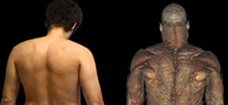
The examiner moves behind the patient and observes:
- Overlying Skin Abnormalities- such as erythema, abrasions, lacerations or scars.
-
The Position of the Scapula: The triangular shaped bone lies over ribs 2 to 7. The Spine of the Scapula is opposite
T3, two inches lateral to the Spinous Process.
Serratus weakness causes "winging" of the Scapula and scoliosis of the Spine may make one shoulder appear lower than the other.
-
Muscle Structure: compare the
bilateral symmetry of the Trapezius, Supraspinatus, Infraspinatus and Posterior Deltoid looking for atrophy or fasciculations.
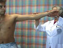
Both Active and Passive Range of Motion are tested. Active Range of Motion is greater than Passive Range of Motion because full active motion of the arm involves motion at the Glenohumeral Joint, combined with motion at the Scapulothoracic articulation. Active range of motion testing is performed independently by the patient. Passive Range of Motion testing is performed by the examiner to assess motion at the Glenohumeral Joint. Motion at the Glenohumeral Joint is isolated by fixing the Scapula with the examiner's hand to prevent Scapulothoracic motion. Examination of a patient with shoulder pain requires comparison of changes in Active Range of Motion and Passive Range of Motion. Range of motion testing at the shoulder includes assessment of:
- Flexion
- Extension
- Abduction
- Adduction
- Internal rotation
- External rotation
Active Range of Motion
Active range of motion (Scapulothoracic and Glenohumeral motion) performed by the patient is assessed by measuring the arc of motion in degrees and noting the smoothness or jerkiness of motion. Active motion is affected by pain, structural changes, joint inflammation, rotator cuff tendonitis, bursitis, and/or weakness. The instructions to the patient are:
- "Move your arm foreward and upward as far as possible." (Forward Flexion up to 180°)
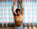
- "Move your arm out away from the side of your body, turn palms up, raise your arm up as far as possible, and touch your hands together over your head." (Abduction up to 180°)
- "Place your hands behind your head." (External Rotation of 40 to 45°)
- "Move your arm back behind your body as far as possible (Extension up to ~45°) and touch your back as close to the shoulder blades as you can." (Internal Rotation of 50-55°)
- "Move your arm in front and across your body as far as possible." (Adduction up to 45°)
Passive Range of Motion
Passive Range of Motion at the Glenohumeral Joint is tested by the examiner. The patient is instructed not to initiate voluntary movement (i.e. "relax, drop your shoulder, let me move your arm"). The examiner grasps the distal upper arm at the elbow and supports the patients' forearm on the examiners' forearm. Care is taken to gently move the patient's arm to the limits of each motion to discern 'end range' characteristics such as severity of pain with motion and whether there is a hard bony end-feel (joint destruction) or a rubbery soft end-feel (tendon or rotator cuff contracture).
Examiner's movements to produce passive shoulder motion are:
-
Internal Rotation:
- With the elbow flexed and at the side of the body, the forearm can be rotated internally ~ 55°.
- With the arm flexed at the elbow and abducted to 90°, the forearm can be rotated downward 80-90°.
-
External Rotation:
- With the elbow flexed and at the side of the body, the forearm can be rotated externally ~ 40-45°.
- With the arm flexed at the elbow and abducted to 90°, the forearm can be rotated up to 90°.
-
Abduction:
- Pull the arm away from the side, raising the elbow until you feel the Scapula start to move (maximum abduction at the Glenohumeral Joint with the Scapula fixed is ~90° and increases up to 120° when the arm is externally rotated).
- Resisted abduction is tested at 30° to isolate Supraspinatus muscle action and at 90° to isolate Deltoid muscle action.
-
Adduction:
- Pull the arm forward and place the hand on the opposite shoulder (~45°).
-
Flexion:
- Move the arm forward up to 90° while preventing Scapular motion.
-
Extension:
- Pull the arm backwards while preventing Scapular motion up to ~45°.
Systematic palpation of the shoulder region will demonstrate abnormalities in relevant anatomical structures and reveal sites of localized tenderness that reproduce the patient's pain.
- Ask the patient to point out the most painful or tender area.
- Carefully palpate the structures in the same order each time to avoid omitting a relevant structure from the exam.
- The technique of palpation should not produce pain itself - palpate with the soft fingertip pad and exert a firm direct pressure, not a circular or 'grinding' motion that may produce pain by shearing and pinching the soft tissues.
- Palpate both the painful and non-painful shoulder for swelling, warmth, effusion, muscle spasm, atrophy and tenderness, comparing one side to the other.
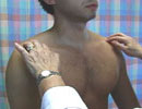
Begin by palpating the
Coracoid Process to determine the patient's responsiveness to the minimally painful pressure of palpation on bony structures. Check the Axillae for Lymphadenopathy. The following structures are palpated:
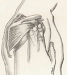
Joint Palpation
Three articulations of the shoulder are highlighted in the transverse sections. Compare each joint bilaterally.
-
Begin with the
Sternoclavicular Joint (S-C), just lateral to the Suprasternal Notch. The Sternoclavicular Joint is the only point of articulation of the shoulder girdle to the trunk, therefore inflammation in the S-C joint will cause increased pain with any shoulder movement.
-
Move your hand laterally along the Clavicle to locate the
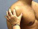
Acromioclavicular Joint (A-C) and ask the patient to "shrug" the shoulder to detect movement at the A-C joint. A-C joint pain may be accentuated by adduction of the arm across the chest and shrugging the shoulders. Note crepitation and/or pain with this movement.
-
The
Glenohumeral Joint (G-H) is palpated 1 cm inferior to the Coracoid Process, which is just below the lateral third of the Clavicle in the
Deltopectoral Groove. With synovitis and/or effusion in the G-H joint, a fluctuant distention may be palpated under the Coracoid Process.
The Rotator Cuff is formed by the conjoined tendinous insertion of the 4 Rotator muscles (Supraspinatus, Infraspinatus, Subscapularis, and Teres Minor). Palpation may be facilitated by pulling the arm into 20 degrees of extension to rotate the cuff anterolaterally from under the Acromion.
-
The superior-lateral cuff is just below the lateral Acromion Process. The insertion of the
Supraspinatus Tendon is at the Greater Tuberosity.
-
Palpate the anterior cuff and the area of insertion of the
Subscapularis Tendon on the Lesser Tuberosity just lateral to the Coracoid Process with the arm positioned in 20 degrees of extension.
-
The Posterior Cuff has the insertions of the
Infraspinatus Tendon on the posterior aspect of the Greater Tubercle, and the
Teres Minor Tendon insertion on the posterior aspect of the Greater Tubercle, just inferior to the insertion of the Infraspinatus Tendon. Palpation of the Posterior Cuff may be facilitated by asking the patient to adduct the arm and place the hand on the opposite shoulder.
Palpate the
Clavicle for protuberances and fractures. The Greater Tuberosity of the
Humerus 1 to 2 cm inferior to the lateral margin of the Acromion allows palpation of the insertion site for the
Supraspinatus Tendon. Occasionally a swollen Subacromial Bursa may be palpated in the indentation between the lateral edge of the Acromion and the Humeral Head. The
Bicipital Groove with the Biceps Tendon is anterior and medial to the Greater Tuberosity between the Greater and Lesser Tuberosity of the Humerus (position arm in External Rotation). Care must be taken with the technique of palpation to use firm direct pressure without a rotating or 'grinding' motion. Excessive palpation pressure on periosteum or the Biceps Tendon in the Groove will produce pain in a normal structure.
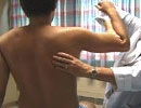
Posteriorly the cervical and thoracic spine should be palpated for tenderness. The Spine of the
Scapula is palpated from the lateral posterior corner of the Acromion moving to the medial border at the level of T3.
A large Subacromial Bursa overlies the Rotator Cuff. A lateral extension of the Bursa is called the Subdeltoid Bursa. An enlarged swollen
Bursa may be palpable under the middle part of the Deltoid, lateral to the Acromion or anteriorly extending from the anterior edge of the Acromion down to the Biciptial Groove. The Subacromial Bursa communicates with the joint cavity in 20% to 33% of individuals past middle age. Any tear of the Supraspinatus Tendon will likely produce a communication between the Bursa and Joint Cavity.
Palpate the muscles bilaterally to compare bulk, consistency and tone and to detect tenderness.
Anteriorly, palpate both heads of the
Pectoralis Major and its insertion on the lateral lip of the Bicipital Groove. The
Biceps Muscles are easier to palpate with the elbow flexed and the arm externally rotated. The Tendon of the Long Head ascends in the Groove and the Short Head ascends to insert on the Coracoid Process. The anterior, middle and posterior parts of the
Deltoid merge and insert at the Deltoid Tuberosity on the lateral mid-Humerus.
Posteriorly palpate the upper (superior)
Trapezius from the Occipital region to the insertion on the Clavicle, Acromion, and the Spine of the Scapula for hematomas or focal tenderness. Palpate the lower Trapezius from the Scapula down to T12. To identify the Rhomboids instruct the patient to place one hand near the back pocket (elbow flexed and arm in internal rotation) and 'push your hand away' to contract the Rhomboids along the medial border of the Scapula. To palpate the
Latissimus Dorsi, ask the patient to abduct the arm and palpate the muscle from the posterior wall of the Axilla to the Iliac Crest. The
Serratus Anterior is palpated as the medial border of the Axilla and extends to the Ribs.