by Adam Lawson BA, MSc and Terra Doucette Hiller BA, BSN, RN
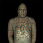
The heart is the muscular portion of the cardiovascular system that rhythmically pumps blood throughout the body. The heart's size for a given individual roughly corresponds to the size of their clenched fist.
The heart consists of four chambers, four valves, and four layers.
Heart Chambers and Valves
There are two types of chambers:
Atria - passively receive blood from the veins at low pressure; they have thinner muscular walls than ventricles.
Ventricles - receive atrial blood and actively pump it through the arteries by contracting their thick muscular walls.
If we follow the flow of blood as it returns to the heart from the veins of the body, it passes through the chambers as follows:
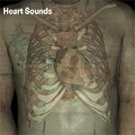
S1 is produced by the simultaneous closure of the mitral and tricuspid valves. Intensity varies with the PR interval.
Examples of underlying pathology include:
Short PR interval - Abnormalities like tachycardia or mitral stenosis can shorten the PR interval. Tachycardia produces greater differences between atrial and ventricular pressures; this pressure difference produces a louder S1. Thickening of the structures due to mitral stenosis can also make S1 louder.
Long PR interval - Abnormalities like a first degree block can lengthen the PR interval and allow the leaflets of the valve to float partially closed before ventricular contraction; the S1 sound becomes softer.
S2 is the closure of the aortic and pulmonic valve. The sound may become louder for several reasons, including: aortic valve stenosis, pulmonic valve stenosis, systemic hypertension, pulmonary hypertension, or a general increase in diastolic pressure.
S3 Heart Sounds (Click to view animation)S3 - indicative of increased ventricular volume and suggestive of heart failure with fluid overload. It is informally known as the "Kentucky gallop" (Ken-tuh-ckey: S1-S2-S3)
A distended or failing ventricle cannot adjust to the increase in fluid. The atrioventricular valve structures (or perhaps the ventricles themselves) vibrate, causing a low-frequency sound best heard with a bell of a stethoscope.
S4 Heart Sounds (Click to view animation)S4 - indicative of atrial contraction in patients with heart failure and decreased ventricular compliance. It is informally known as the "Tennessee gallop" (Ten-nes-see: S4-S1-S2)
The sound may be caused by: hypertension, myocardial infarction, angina, cardiomyopathy and aortic stenosis.
Atrial contractions forcing blood into a highly resistant, non-compliant ventricle produce the S4 sound. It is best heard using the bell at the apex of the heart.
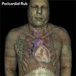
Cardiac motion occurring within an inflamed pericardial sac produces a distinctive and audible sound which can be accentuated by having the patient lean forward and exhale. It can be heard in cardiac tamponade.
The cardiac muscle of the left ventricle supplies blood to the body through a vast smooth muscle network of arteries before draining it back to the heart through an often parallel system of veins. Smooth muscle allows vessels to contract, expand, and facilitate the pumping of blood.
Superficial veins are not paired with an artery and run close to the body surface to help regulate temperature. Their number and their proximity to the skin surface also makes them easy to harvest for several surgical procedures.
Due to the lower pressures of venous blood, veins have thinner walls and valves to prevent backflow.
The branches of aorta supply blood to the entire body. It emerges from the heart and ascends briefly before curving to descend through the thorax, abdomen, and pelvis. It is subdivided into several sections that each have their own branches:
Ascending aorta - the shortest portion of the aorta emerging from the left ventricle that carries blood in the superior direction.
Descending aorta - the remainder of the aorta that carries blood in the inferior direction to supply the trunk and lower limbs.
Thoracic aorta - the continuation of the aortic arch which ends at the diaphragm. It has small, bilateral branches at the upper rib levels that supply the chest wall and branches for mediastinal structures such as the esophagus and trachea.
Abdominal aorta - the continuation of the thoracic aorta, it supplies the organs of the abdomen and pelvis. Major abdominal aortic branches, listed in order of their occurrence, include:
Celiac trunk - supplies several other important branches:
External iliac arteries - travel deep to the inguinal canal where their name changes to the femoral arteries. The femoral arteries travel the length of the thigh and are renamed the popliteal arteries in the posterior knee. Entering the leg, the popliteal arteries have two major branches: