Anatomy Relevant to Acute Respiratory Distress Syndrome
by Adam Lawson BA, MSc and Terra Doucette Hiller BA, BSN, RN
Nervous System Control of Breathing
Ventilation control is not completely understood because of its complexity. The brain, muscles, chemoreceptors, mechanoreceptors, and nerve fibers all play a role in ventilation regulation.
-
Brain Regulation
- Cerebral cortex - allows voluntary ventilation
-
Brainstem - regulates automatic ventilation. Ventilation depends on the rhythmic function signaled by the brainstem and its intact nervous pathways to the respiratory muscles.
-
Medulla oblongata - sends signals through the phrenic nerves which initiate the diaphragm contraction.
- Dorsal respiratory group - controls the basic rhythm of ventilation; the cells will automatically fire and trigger inhalation.
- Ventral respiratory group - controls voluntary or increased rates inspiration and expiration.
-
Pons - modulates ventilatory patterns and coordinates transition from inhalation to exhalation.
- Pneumotaxic center - controls rate and pattern of respiration by limiting inhalation and triggering exhalation.
- Apneustic center - controls depth of inspiration by working antagonistically with limiting signals of the pneumotaxic center.
-
Medulla oblongata - sends signals through the phrenic nerves which initiate the diaphragm contraction.
-
Chemoreceptors - regulate ventilation by detecting chemical changes in blood and other fluid and sending signals to the cerebral cortex. There are two types of chemoreceptors:
-
Central chemoreceptors - located on the ventrolateral surface of the medulla.
- Indirectly responsive to changes in PaCO2.
-
Peripheral chemoreceptors - send signals to the respiratory control centers to immediately regulate breathing and blood pressure.
- Carotid body - located at the bifurcation of the common carotid artery. They are responsive to changes in oxygen levels, CO2 levels, pH, and temperature; sends signals via the glossopharyngeal nerve.
- Aortic body - located in the aortic arch between the brachiocephalic trunk and the left common carotid artery. They are responsive to changes in oxygen and CO2 levels; sends signals via the vagus nerve.
-
Central chemoreceptors - located on the ventrolateral surface of the medulla.
-
Spinal cord - processes information and conducts signals from the brain and peripheral receptors to the muscles of ventilation.
- The phrenic nerve, which innervates the diaphragm, is formed from fibers of the C3, C4, and C5 spinal nerves.
- Intercostal nerves originate between the T1 and T12 vertebrae and innervate the intercostal muscles.
-
Miscellaneous Lung Receptors
- Irritant receptors - lie in the epithelial cells of the bronchioles. They respond to irritants by inducing bronchoconstriction and rapid, shallow breathing (i.e., tachypnea).
- Stretch receptors - located along the entire airway. Changes in lung volume cause them to inhibit inhalation and prevent lung over inflation by altering respiratory rate and tidal volume.
- J receptors - lie in the alveolar walls close to the capillaries. They respond to engorgement of the pulmonary capillaries and an increase interstitial fluid volume by inducing rapid, shallow breathing.
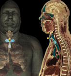
Lymphatic Circulation
Lymphatic tissue within and around the lungs protects ventilatory pathways to the alveoli by facilitating cell- and antibody- mediated immune response and by clearing the bronchi of excess fluid. The lungs, when compared with other organs, are associated with the greatest quantity of lymphatic tissue.
- Afferent lymphatic vessels are first found around and above the level of the terminal and respiratory bronchioles.
- Vessels from the bronchioles converge at the bronchopulmonary nodes surrounding the bronchi of the lungs.
- Lymph travels upward from the bronchopulmonary nodes to the tracheobronchial nodes or the paratracheal nodes.
- Lymph eventually drains into the left thoracic duct and flows back into blood circulation when the duct drains into the left subclavian vein.
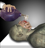
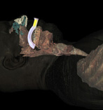
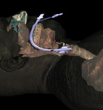
Anatomy involved during Intubation
Several methods can be used to help the airway function of a compromised patient:
- Bag valve mask (a.k.a., BVM, Ambu bag) - a mixture of air and oxygen is forced from a bag into the patient's lungs through a valved mask covering the nose and mouth.
-
Oropharyngeal airway (a.k.a. oral airway) - helps to prevent the tongue from covering the epiglottis and airway.
- An oral airway should only be used with unconscious patients.
- Typically, the length of the tube will run from the corner of the mouth to the tip of the ear lobe.
- To place the tube, insert it with the J pointing upward toward the rood of the mouth. When the tube is halfway in, twist the tube 180° so the J points down toward the trachea and finish insertion.
-
Endotracheal tube (a.k.a., ET tube, ETT) - inserted through the mouth, larynx, and vocal cords into the trachea of an unconscious patient.
- Typically performed by a physician.
- The ET tube may also be placed down a patients nose; this is called nasoendotracheal intubation.
-
Tracheostomy - insertion of a specially-designed tube directly into the trachea through an tracheotomy incision. The tube is typically inserted through:
- A midline incision on the neck between the infrahyoid muscles.
- An incision made on a region of the trachea in between the second and third tracheal rings known as the Björk flap.
(Click to view animation)
(Click to view animation)
(Click to view animation)
(Click to view animation)