Anatomy Relevant to Chest Tube Insertion
by Adam Lawson BA, MSc and Terra Doucette Hiller BA, BSN, RN
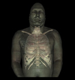
Pulmonary assessment should start superiorly at the upper respiratory system and move inferior toward the intersection of the respiratory system with the cardiovascular system.
Upper Respiratory System
-
Nasal cavity - there are five bones which form most of the enclosing surface of the nasal cavity:
- Nasal bone
- Maxillary bone (a.k.a., maxilla)
- Ethmoid bone
- Sphenoid bone
- Frontal bone
- Paranasal sinuses - four bilateral sinuses:
-
Pharynx - subdivided into the:
-
Nasopharynx - the superior portion of the pharynx which connects to the posterior portion of the nasal cavity via the internal nares.
- It provides the primary route for inspired air.
- The soft palate separates it from the oral cavity.
-
Oropharynx - the portion of the pharynx which extends from the posterior oral cavity to about the level of the hyoid bone.
- It provides an alternative or supplementary route for inspired air when respiration increases or when the nasopharynx is congested.
- Laryngopharynx - the inferior portion of the pharynx which communicates with the trachea (via the larynx) and esophagus.
-
Nasopharynx - the superior portion of the pharynx which connects to the posterior portion of the nasal cavity via the internal nares.
Lower Respiratory System
-
Larynx - connects the pharynx to the trachea.
- It produces vocal sound with a variety of structures including the hyoid bone and vocal cords.
- It prevents aspiration of particles by covering the tracheal opening with the epiglottis during swallowing.
-
Trachea - a cartilaginous tube that begins superiorly at about the same level as the C6 vertebra and ends inferiorly at about the level of the T5.
- It allows passage of air to and from the pharynx and bronchi of the lungs.
- 15 to 20 imperfect cartilaginous rings stiffen the tracheal wall and help protect the airway from traumatic injury, collapse, or overexpansion during transient changes in airway pressure.
-
Carina - the cartilaginous ridge at the bifurcation of the trachea into the primary bronchi.
- Sensory neurons innervate the carina; aspirant which stimulates these neurons will initiate a cough reflex and bronchoconstriction.
-
Primary Bronchi - The trachea divides into the right and left bronchus before entering the lung.
- The left bronchus is longer and more narrow than the right; it projects at about 45° to 50° from the midline.
- The right bronchus projects about 20° to 30° from the midline.
-
Bronchi - The division of the trachea into the left and right bronchi is the primary (a.k.a., first, principal) "generation" of bronchi; there are up to 18 further generations of bronchi, later generations having smaller diameters than those preceding.
- Cartilaginous rings are found in the trachea and, with decreasing regularity, in the first three generations of bronchi; subsequent generations (i.e., bronchioles) have walls composed of smooth muscle.
- Bronchioles - the remaining 14 generations of bronchi; they are categorized based on which lobe they supply with air.
-
Lungs - the organs of respiration.
- The right lung has more lobes and is bigger than the left lung because of the of the heart's position.
- The left lung is longer than the right because the large size of the liver beneath the diaphragm reduces the space available for right lobe of the lung. The right lung ends at about rib five while the left lung ends at about rib six.
- Only 3 of the 5 lobes of the lung can be auscultated from the anterior thorax; the:
- Auscultate from the posterior thorax the:
Lung Auscultation
(Click to view animation)
Chest tube insertion sites
There is approximately 5mL of fluid between the parietal and visceral pleura which help to decrease friction during respiration. The lymphatic system can drain up to 500mL of excess fluid production per day, but production exceeding that amount will cause fluid to built up within the pleural cavity.
Accumulation of air, blood, or fluid within the pleural cavity will reduce the negative pressure in the pleura required for normal respiration; a chest tube may be used to drain this excess.
Density of the accumulation determines the placement of the chest tube; for example, the insertion site for a chest tube removing air would be higher than the site for a tube removing fluid.
Indications for a chest tube include:
-
Pneumothorax - accumulation of air in the pleural space
- Common causes - trauma, lung disease, invasive thoracic procedures, or forceful coughing.
- Placement - near the second intercostal space along the midclavicular line of the affected side.
-
Hemothorax - accumulation of blood in the pleural space
- Common causes - blunt trauma, penetrating trauma, or a complication of thoracic surgery.
-
Placement - at the mid or anterior axillary line of the affected side. Typically the tube is placed:
- Behind pectoralis major - to avoid difficult penetration of the thick muscle.
- Above the 5th rib - to avoid interference from the diaphragm as it rises to this level.
-
Pleural effusion- accumulation of fluid in the pleural space.
- Common causes - left ventricular heart failure, pulmonary embolism, pneumonia, cancer, or interference of the lymphatic system.
-
Chylothorax - accumulation of lymphatic fluid in the pleural space.
- Common causes - chest trauma, tumor, or a complication of surgery.
-
Empyema - accumulation of pus in the pleural space
- Common causes - bacterial pneumonia, thoracic trauma, rupture of a lung abscess, abdominal infection, or esophageal tearing.
- Drainage may also be required for post-cardiac surgeries that involve opening the heart such as: valve replacement, CABG or heart transplantations. Placement into the mediastinum drains blood around the heart. The chest tube is often placed while the chest is still open and the chest is closed around the tube.