Organs of the Gastrointestinal and Urinary Systems
Most of the gastrointestinal organs are part of the alimentary canal. They can be found in the following order as the canal is followed from beginning to end: (Figure 3-5)
- esophagus
- stomach
- small intestine
- large intestine
- rectum
- anal canal
Other organs are associated structurally and functionally with the alimentary canal but are not a direct part of it. These organs include:
- liver
- gallbladder
- pancreas
The spleen, stomach and intestines have common blood vessels, however unrelated functions keep the spleen from being grouped with the gastrointestinal organs. Kidneys, ureters and urinary bladder are not directly related to the G.I. system.
Remember, the alimentary canal is a long tube, (±25 feet). Different stages of digestion and absorption of food occur along the entire length.
The esophagus extends from the pharnyx to the stomach, a distance of 9 to 9.5 inches (23 to 25 cm). It empties into the stomach directly inferior to the diaphragm through the cardiac orifice.
The stomach is situated in the left upper quadrant of the abdomen. The size, shape and general location vary greatly depending on the contents and their stage of digestion. It empties into the duodenum through the pyloric sphincter.
The duodenum is the first and shortest part of the small intestine. It extends in an almost circular course so its termination point is not far from its beginning point. The length of 9.5 to 10 inches (25 cm) gives it its name. It is equal in length to the breadth of twelve fingers. It receives digestive enzyme secretions from the pancreas and bile from the gallbladder.
The remainder of the small intestine is made up of the jejunum and ileum. The jejunum makes up the first two fifths and the ileum the last three fifths. The jejunum and ileum absorb most of the nutrients from the food passing through the alimentary canal.
The ileum empties into the large intestine at the ileocecal junction. This is so named because the first part of the large intestine is a blind pouch called the cecum. The appendix, a small blind tube itself, is found arising from the cecum.
The large intestines "frame" the small intestines on three sides. So on the right side there is an ascending colon, superiorly, a transverse colon, and on the left side a descending colon and sigmoid colon. The sigmoid colon is continuous with the rectum and anal canal in the pelvis.
The liver lies in the most superior upper right quadrant of the abdomen just inferior to the diaphragm. The left lobe extends part way into the upper left quadrant. It is very important in assisting the digestive system in many ways. The venous hepatic portal blood system delivers venous blood from the entire digestive system to the liver. The blood in the hepatic portal vessels carries vital nutrients and some occasional toxins. Multiple metabolic processes in the liver act on the nutrients to transform them into materials the cells of the body can utilize.
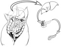
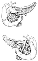
The gallbladder is a small sac that concentrates and stores bile produced in the liver. The gallbladder, tucked under the right lobe of the liver, receives bile from each of the liver lobes via hepatic bile ducts. Bile is a natural detergent utilized by the G.I. system to digest fat. When a meal high in fat is consumed, the gallbladder contracts and forces bile into the cystic duct which is continuous with the common bile duct. The common bile duct empties into the duodenum. (Figure 3-6)
The pancreas is held in the hair-pin curve of the duodenum and it has endocrine and exocrine glandular functions. It is an endocrine gland, secreting insulin into the blood stream, and an exocrine gland, producing digestive enzymes that empty via the common bile duct into the duodenum. (Figure 3-6)
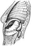
The spleen is the largest lymph organ in our body and not only carries out lymph functions but also breaks down blood platelets and old red blood cells. It is not directly related to the G.I. system but its venous return empties into the liver like the G.I. organs. This unique organ is located in the left superior quadrant of the abdomen, just under the lower left ribs. (Figure 3-7)
The kidneys are located retroperitoneally on either side of the posterior wall of the abdomen, slightly inferior to the liver. They function as blood "filters" and have large renal arteries entering them from the aorta, and large renal veins emptying into the inferior vena cava.
Urine, the filtrate of the kidneys, exits the kidneys via the ureters, which descend from the hilus retroperitoneally along the posterior wall of the abdomen and pelvis to empty into the urinary bladder. (Figure 3-7)