Thoracic & Abdominal Vasculature
Directions
- Print this PDF worksheet for a hardcopy guide as you work through this lesson.
- Within the lesson click the red linked headings to bring up the desired starting point within the cadaver for your work.
- Use the provided images on the worksheet to annotate and identify specific anatomical structures.
This dissection investigates the major blood vessels in the thoracic and abdominal regions. To begin, rotate your specimen to gain a posterior perspective. Next, locate each of the following:
- Aortic Arch (This better be easy since you were supposed to master the aortic arch in the previous dissection.)
- Thoracic Aorta
At about 8 in. (20 cm) long, you see that the thoracic aorta is a continuation of the aortic arch. It begins between the fourth and fifth thoracic vertebrae where it lies to the left of the vertebral column. As it descends, it moves closer to midline and extends through the diaphragm. Many visceral and parietal arteries branch off the thoracic aorta. Visceral arteries provide blood to various organs while the parietal branches provide blood to body wall structures. As an example, locate the following:
- Posterior Intercostal Arteries - These supply blood to muscles as well as other more superficial structures and the spinal cord/meninges. Intercostal arteries are "parietal" branches from the thoracic aorta.
Locate the:
- Abdominal Aorta - The abdominal aorta is a continuation of the thoracic aorta. It begins where the aorta passes through the diaphragm.
Notice the diaphragm is now shown in this posterior view. This gives you a good idea of how/where the aorta passes through. Before we continue with our cardiovascular examination, fade out the ghosted external anatomy. Next dissect away the diaphragm.
Like the thoracic aorta, the abdominal aorta gives off visceral and parietal branches. Unpaired branches arise from the anterior aortic surface, so use the rotation tool to change your perspective to an anterior view.
Select the "Index" tab and type in "Celiac Trunk". Select it from the list and click the "Add & Highlight" button at the bottom. Once its highlighted in red you'll quickly see why I didn't want you searching for this little guy. It is the first visceral branch from the aorta inferior to the diaphragm. The reason it is so small is because it divides almost immediately into three branches. Identify each of these celiac trunk branches:
- Left Gastric Artery - The left gastric artery is the smallest of the three branches. It passes superiorly from the celiac trunk and drifts to the left (anatomical left) toward the esophagus and then turns to follow the lesser curvature of the stomach. It provides blood for the stomach and esophagus.
- Splenic Artery - This is the largest branch of the celiac trunk. As you can see, it arises from the left side of the celiac trunk distal to the left gastric artery. Once it arrives at the spleen it subdivides into three smaller arteries.
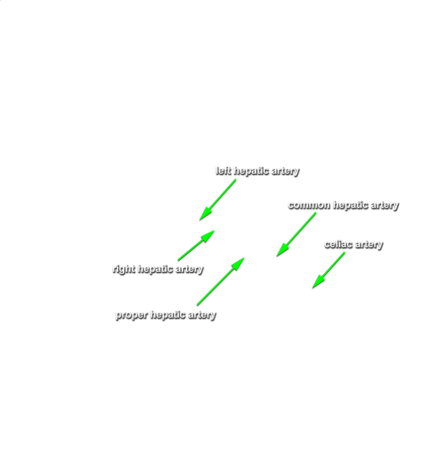
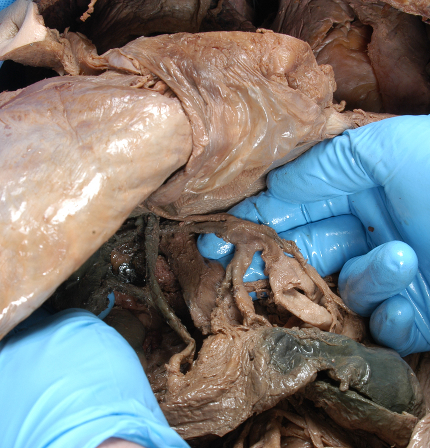
- Common Hepatic Artery - The common hepatic artery is intermediate in size of the three arteries that arise from the celiac trunk. Unlike the other two, however, the common hepatic artery arises from the right side of the celiac trunk. It also gives rise to three smaller arteries.
The next two arteries branch off the abdominal aorta a bit inferior to the celiac trunk.
- Superior Mesenteric Artery - About a centimeter inferior to the celiac trunk, the superior mesenteric artery branches from the abdominal aorta. It give rise to 5 smaller arteries that supply blood to the intestine and colon.
- Inferior Mesenteric Artery - About 6 cm further down the anterior surface of the abdominal aorta, the inferior mesenteric artery branches off and supplies blood to the colon through three smaller branches.
Locate the following two PAIRED visceral arteries that branch off the lateral abdominal aorta:
- Renal Arteries (Right & Left) - These arteries branch off the lateral abdominal aorta about a centimeter inferior to the superior mesenteric artery. Note how the right renal artery is longer than the left and emerges slightly lower. These arteries carry blood to the kidneys, adrenal glands, and ureters.
-
Gonadal Arteries (Right & Left) - The gonadal arteries typically arise about a centimeter inferior to the renal arteries and provide blood to their respective reproductive organs.
As you have just discovered, the arterial system delivers oxygenated blood to visceral and parietal destinations in the thorax, abdomen, and pelvis. We obviously need to get this blood circulated back to the heart which is the job of the venous system. Arteries subdivide to form arterioles which subdivide to form capillaries that provide circulatory services to body tissues. Capillaries flow together to form venules which combine to form veins. We'll now discover some primary veins and follow them as they return blood to the right atrium of the heart.
As soon as you are oriented to the portion of the body we are examining, use the right slider to fade out the external anatomy so you have a better view of these veins. Since we are returning blood to the heart, let's start our investigation with inferior vessels conducting blood distally from the heart.
- Internal Iliac Veins (Right & Left) - These veins receive blood returning from the legs and external genital regions.
- External Iliac Veins (Right & Left) - These veins drain blood from the lower limbs and abdominal wall.
- Common Iliac Veins (Right & Left) - This large vein is formed from the union of the internal and external iliac veins.
- Inferior Vena Cava - The two common iliac veins unite to form the inferior vena cava which is the largest vein in the body. It is about 3.5 cm. (1.4 in.) in diameter. It receives blood from the entire lower half of the body. Some of you may have experienced edema (swelling) of the ankles and feet and even varicose veins during the later stages of pregnancy. This is due to compression of the inferior vena cava thereby reducing the body's ability to drain blood and interstitial fluid from the lower half of the body.
- Renal Veins - These veins drain blood from the kidneys and empty into the inferior vena cava.
- Suprarenal Veins - These veins drain blood from the adrenal (suprarenal) glands. Notice that the left suprarenal vein empties into the left renal vein while the right suprarenal vein empties into the inferior vena cava.
- Testicular Veins - Gonadal veins are obviously gender specific, testicular in males and ovarian in females. As you would expect, these veins drain blood from their respective reproductive organs. Note that the left testicular vein empties into the left renal vein whereas the right testicular vein empties directly into the inferior vena cava. Ovarian veins follow this same pattern.
- Hemiazygos Vein - The hemiazygos vein receives blood from the left posterior intercostal veins and the accessory hemiazygos vein.
- Accessory Hemiazygos Vein - See how the left intercostal veins just seem to empty into nothing. Obviously, that's not the case. They empty into the accessory hemiazygos vein which, for some reason, is not present and identified in your specimen. We'll chalk it up to a "thready" vessel that didn't survive the dissection process. The accessory hemiazygos vein receives blood from left intercostal veins draining the left thoracic wall.
- Azygos Vein - The azygos vein receives blood from the hemiazygos and accessory hemiazygos (if the accessory hemiazygos vein doesn't empty directly into the hemiazygos), right subcostal and right ascending lumbar veins. The azygos vein empties into the SUPERIOR vena cava.
Here's a nice little review for you. Most of the vasculature identified thus far is seen here. Let's see how you do identifying it all. :-o
And finally, let's take a look at blood circulation through the liver. A portal vein is one that carries blood BETWEEN capillary systems. Thus far you have not seen anything like this occurring. Typically, blood moves from arteries to arterioles to capillaries to venules to veins and back to the heart.
We haven't studied the digestive system as yet so you'll be tracking through some unlearned anatomy to get at the hepatic portal venous system. I want you to start dissecting away the small intestine one section at a time starting with the most inferior 18th segment of the ileum. Work carefully and remove all segments of the ileum, jejunum, and the duodenum. Next, dissect the stomach, pyloric orifice, liver and the spleen. If I've covered everything I believe that leaves you with only the hepatic portal system.
- Splenic Vein - The splenic vein drains blood from capillaries of the spleen, parts of the stomach and portions of the large intestine.
- Superior Mesenteric Vein - This vein drains capillary blood from the small intestine, colon, and parts of the stomach.
- Inferior Mesenteric Vein - This vein drains distal large intestine tissues and the rectum. It joins the splenic vein just before that vessel unites with the superior mesenteric vein to form the hepatic portal vein.
- Gastric Vein - This vein drains the stomach tissues.
-
Hepatic Portal Vein - This vein forms with the union of the splenic and superior mesenteric veins.
- Right & Left Branches - These subdivide to form the capillaries that deliver oxygen poor yet nutrient rich blood to the sinusoids of the liver. Some nutrient molecules are stored there while others are modified and return to general circulation. Sinusoids are large, thin-walled, and rather leaky capillaries. As an example of nutrient molecule modification, blood sugar levels can be lowered as monosaccharide glucose molecules are converted to polysaccharide glycogen. The liver also detoxifies the blood. Alcohol is removed as are many bacteria.
- Hepatic Veins - Eventually, blood leaves the liver sinusoids through the hepatic veins which drain into the inferior vena cava.
- Hepatic Artery (identified earlier) - As you investigate this anatomy you'll rediscover the hepatic artery (a branch of the celiac trunk) which supplies freshly oxygenated blood that mixes with the deoxygenated blood within the liver sinusoids.
And with that we are done with the gut, so to speak, and ready for your final adventure down vascular lane with an examination of arteries and veins of the lower limbs.
Self-test Labeling Exercises