


You're obviously looking at the external anatomy. Reveal the cranial nerves by fading out the external anatomy using the skin control in the toolbar below your dissection.
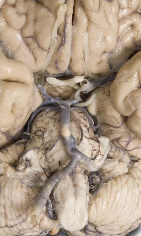
There are twelve pairs of cranial nerves that pass through various foramina in the cranial bones. Cranial nerves are part of the peripheral nervous system. The best way to identify these nerves is by using the "Systems" tab. Click that tab, and expand each of the following; "Nervous system", "Peripheral nervous system" and "Cranial nerves". All of the cranial nerves are already visible in your dissection so as we walk through each one in this lesson you will be using the systems tab to highlight them.
Cranial Nerve I: Olfactory Nerve (CNI) - The olfactory bulb and tracts are already highlighted for you. The olfactory nerve is one of the two purely sensory cranial nerves. It conducts nerve impulses for the sense of smell (olfaction). Rotate your specimen to the lateral perspective. By simply mousing over the highlighted CNI, identify the olfactory bulbs and tracts. These are actually separate structures but are labeled together in your specimen. Within the olfactory bulbs, the axon terminals of olfactory receptors form synapses with the dendrites and cell bodies of the next neurons in the olfactory pathway. The axons of these neurons make up the olfactory tracts which extend posteriorly from the olfactory bulbs. Axons in the olfactory tracts end in the primary olfactory area in the temporal lobe of the cerebral cortex.
Cranial Nerve II: Optic Nerve (CNII) - Rotate your specimen back to the anterior perspective. Click any black space in the dissection area to de-highlight CNI. Return to the systems tab to highlight the the optic nerve (CNII) by selecting it and clicking the "Add & Highlight" button. The optic nerve is the second entirely sensory cranial nerve which contains axons that conduct nerve impulses for vision. Retinal rods and cones in each eye initiate visual signals relaying them to bipolar cells that pass the signals on to ganglion cells that join to form the optic nerve from each eye.
Identify the optic chiasm (KI-aszm = a crossover as in the letter "X") by simply mousing over parts of CNII. As you see (no pun intended), this is the center of CNII where the medial optic nerve axons from each eye cross to the opposite side. Axons from the lateral half remain on the same side. Because the lens system of each eye reverses all images, the medial half of each retina receives light rays from the temporal part of the visual field (that is, from the far left or far right rather than from straight ahead), and the lateral half of each retina receives an image of the nasal (central) part of the visual field. In this way, the right and left optic tracts carry and send a complete representation of each half of the visual field.
Cranial Nerve III: Oculomotor Nerve (CNIII) - De-highlight CNII and again use the systems tab to highlight the oculomotor nerve (CNIII). The oculomotor nerve is mainly motor in function but is classified as a "mixed" nerve since it has some sensory function, too. The primary function is muscular control of the eyeball and upper eyelid. Sensory function includes proprioception (position and movement).
Cranial Nerve IV: Trochlear Nerve (CNIV) - De-highlight CNIII and highlight the trochlear nerve (CNIV). As you can tell from the dissection, the trochlear nerve is the smallest of the 12 cranial nerves. It is a "mixed" nerve and primarily motor. Similar to the oculomotor nerve, the trochlear nerve innervates an eye muscle. Proprioceptive functions are similar to the oculomotor nerve, too, in that eye position and movement information is carried to the midbrain.
Cranial Nerve V: Trigeminal Nerve (CNV) - The trigeminal is the largest of the cranial nerves and is clearly mixed in function. Use highlighting once again, to locate this nerve. By "mousing over" the dissection, locate the three branches of the trigeminal nerve.
- Ophthalmic - The smallest branch, it carries sensory impulses from the skin over the upper eyelid, eyeball, lacrimal glands, upper part of the nasal cavity, side of the nose, forehead, and anterior half of the scalp.
- Maxillary - Intermediate in size, it carries sensory impulses from the nose mucosa, palate, parts of the pharynx, upper teeth, upper lip, and lower eyelid.
- Mandibular - The largest branch, it is composed of sensory axons from the anterior two-thirds of the tongue (not taste), cheek and mucosa deep to it, lower teeth, skin over the mandible and side of the head anterior to the ear and the mucosa of the mouth floor. It also contains somatic motor axons that supply muscles of mastication.
Cranial Nerve VI: Abducens Nerve (CNVI) - The abducens is mainly a motor cranial nerve BUT it is technically classified as "mixed". As before, highlight it so that it stands out in the dissection. The name of this nerve derives from its function of "abducting" the eyeball by lateral rotation through its innervation of the lateral rectus muscle. Sensory axons extend from proprioceptors located in the lateral rectus muscles.
Cranial Nerve VII: Facial Nerve (CNVII) - The facial nerve is a mixed nerve, too, with sensory axons extending from the tastes buds located in the anterior two-thirds of the tongue as well as proprioceptors in the facial and scalp muscles. Somatic motor neurons innervate muscles of the face plus the stylohyoid muscle and digastric. Parasympathetic neurons extend to the lacrimal glands (that secrete tears), nasal glands, palatine glands, and saliva producing sublingual and submandibular glands. If you haven't done so already, locate this nerve using highlighting as before.
Cranial Nerve VIII: Vestibulocochlear Nerve (CNVIII) - The vestibulocochlear nerve is nearly entirely sensory and was formerly known as the acoustic or auditory nerve. The vestibular branch carries impulses for equilibrium (from the semicircular canals) whereas the cochlear nerve branch carries impulses for hearing (from the cochlea). Again, use highlighting to identify it in your dissection.
Cranial Nerve IX: Glossopharyngeal Nerve (CNIX) - The glossopharyngeal nerve is mixed with both sensory and motor neurons. Sensory neurons come from the taste buds, somatic sensory receptors on the posterior third of the tongue, swallowing proprioceptors, and carotid stretch and chemoreceptors. Motor neurons innervate the stylopharyngeus, which is partially responsible for swallowing. If you haven't already done so, "Add & Highlight" to locate the glossopharyngeal nerve.
Cranial Nerve X: Vagus Nerve (CNX) - Vagus and "vagrant" come from the same Latin root meaning "wandering." A sensory and motor mixed nerve, the vagus is distributed from the head and neck into the thorax and abdomen. Sensory functions include touch from the external ear, a few taste buds, neck and throat proprioceptors, carotid and aortic stretch and chemoreceptors, as well as visceral sensory receptors in most organs of the thoracic and abdominal cavities. Parasympathetic motor nerves supply gastrointestinal glands and smooth muscle of the respiratory passageways, esophagus, stomach, gallbladder, small intestine, and most of the large intestine. Locate this cranial nerve by highlighting it in your dissection.
Cranial Nerve XI: Accessory Nerve (CNXI) - The accessory nerve is another mixed cranial nerve. It is unique, however, in that it originates from both the brain stem and the spinal cord. The cranial root is motor and innervates voluntary swallowing muscles. The spinal root is mixed but mainly motor with axons arising in the anterior gray horn of the first five segments of the cervical portion of the spinal cord. These axons conduct impulses that coordinate head movements (sternocleidomastoid and trapezius muscles). Sensory axons originate from proprioceptors in these muscles. As before, highlight it for identification.
Cranial Nerve XII: Hypoglossal Nerve (CNXII) - The last of our 12 cranial nerves, the hypoglossal is another mixed nerve. The sensory axons originate from tongue proprioceptors (now you know what to blame when you bite your tongue) while motor neurons conduct impulses to tongue muscles.