As you see, the respiratory system returns us to a more centralized set of organized organs.
We'll begin this excursion with inhalation. Air is warmed and cleansed after it enters the respiratory system through the external nares (nostrils). Air enters the nasal cavity, which is divided into lateral halves.
The nasal cavity and mouth are connected to the larynx and esophagus by the pharynx, commonly called the throat. Extending about 13 cm, the pharynx has three regions. Locate the following pharyngeal subdivisions.
The pharynx is the passageway for both air and food. Locate the pharyngeal tonsils, you learned about them back in the lymphatic system, they compose part of the posterior wall of the nasopharynx. As the nasopharynx blends into the oropharynx, the epithelium lining the pharynx changes from pseudostratified columnar epithelium to a more protective stratified squamous epithelium. This structural adaptation reflects the increased friction and greater chemical trauma accompanying food passage.
The epiglottis is elastic cartilage that covers the opening to the larynx when you swallow, temporarily stopping breathing and giving food the "right of way" (a critical role since you want dinner to end up in your stomach and not in your lungs). If anything other than air enters the larynx, the cough reflex is initiated, which acts to expel the unwanted substance. This protective reflex does not work when we are unconscious, so it is never a good idea to administer fluids when you're attempting to revive an unconscious person.
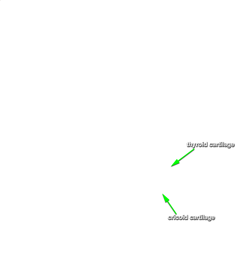
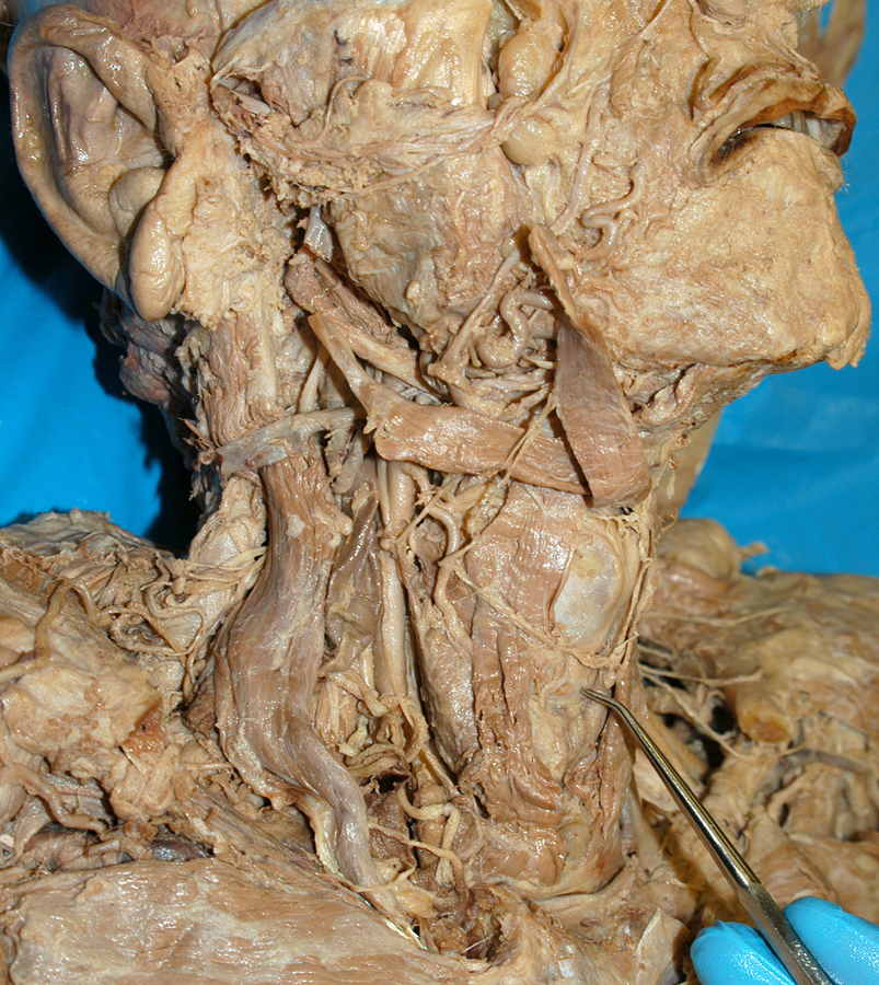
The larynx includes nine cartilaginous structures, one is the epiglottis. Two of the remaining eight will be examined along with two structures that directly produce our voice.
Dissect away the thyroid cartilage.
The superior portion of the larynx is lined with stratified squamous epithelium where it is subject to food contact. Pseudostratified ciliated columnar epithelium lines the inferior portion. The cilia move mucus upward toward the pharynx and away from the lungs. We help this movement when we "clear our throats."
We're going to reverse our direction of study for this segment and examine the lungs. I'm pretty certain you don't need this course to know that inhaled air ends up in our lungs! :-o You probably don't know, however, that our two lungs are asymmetric. Investigate this asymmetry in your specimen. You'll discover that each lung is composed of lobes and herein lies the bilateral difference. (Fade down the skin opacity for better lung resolution).
A horizontal fissure separates the right superior lobe from the middle lobe. An oblique fissure separates the middle lobe from the right inferior lobe. The left lung has no middle lobe so an oblique fissure separates the superior from the inferior lobes.
It's not obvious in your specimen, however, the lung surface has a distinctive texture. Small hexagonal shapes range from about the size of a pencil eraser to the size of a penny. These are the smallest subdivisions of the lung that are visible with the naked eye. They are called lobules.
Dissect away all of the lobes of the lung to reveal the internal airways. The branching you have revealed is called the bronchial tree.
Suffocation is possible when someone has a piece of food lodged in the trachea or the glottis of the larynx. The Heimlich maneuver is a procedure in which the air in a person's lungs is used to force out an obstructing piece of food or other matter from the windpipe.
Note that the right lung (with its three lobes) has three lobar bronchi (go figure, eh?) while the left lung has only two. You need to rotate your specimen to really see the dimensionality.
The segmental bronchi further divide to form even smaller bronchioles which subdivide even further to form terminal bronchioles. Neither of these are detailed in your specimen due to their size and delicate nature. Mucus-producing cells are absent from the bronchioles. Any remaining debris in the inhaled air is removed by macrophages located in the lung alveoli (air sacs) at the end of the terminal bronchioles.
Well, wasn't that a breath of fresh air! :-)