Identify the structures associated with the cavernous sinus.
- (ON THE SIDE WITHOUT THE BRAIN) Use the hand electric saw to make a horizontal cut through the cranium. Start the cut at the midline approximately 1 cm superior to the orbital margin. Continue the cut in the posterior direction ending along the midline approximately 1 cm superior to the external occipital protuberance. You may need to remove a portion of the temporalis muscle to facilitate the cut. Pry the dura away from the bone and remove the calvarium.
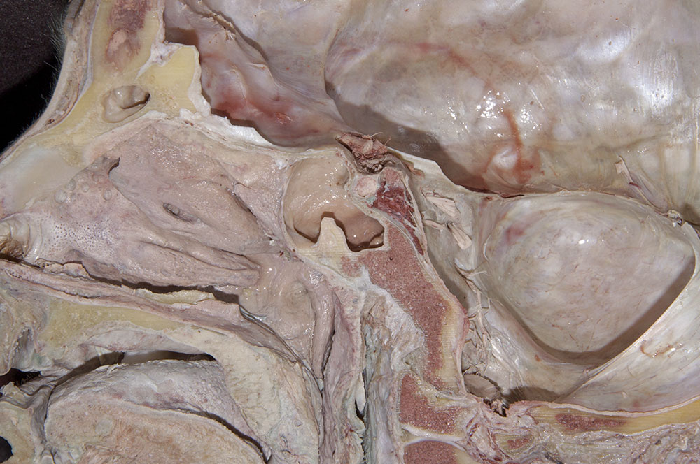
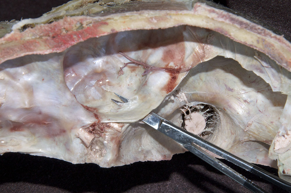
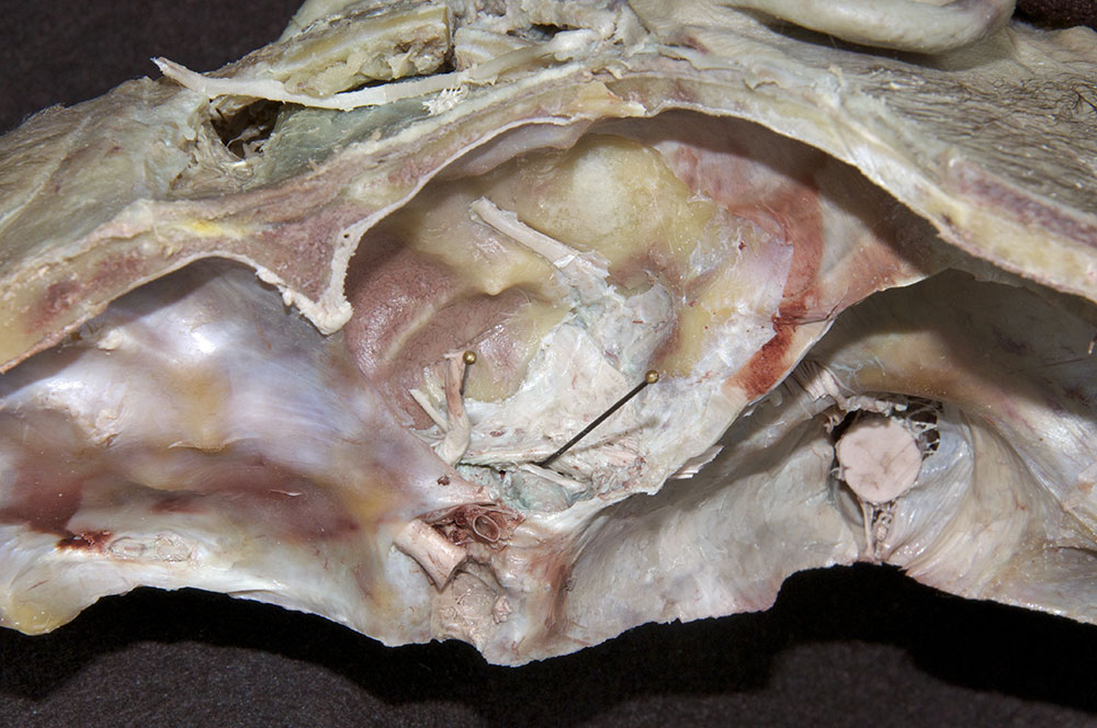
- (ON THE SIDE WITHOUT THE BRAIN) Identify the pituitary (hypophysis), sellar diaphragm and cavernous sinus. (G 7.30A;N 105;Gl 40.18)
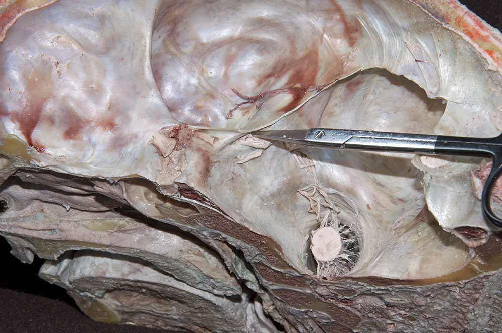
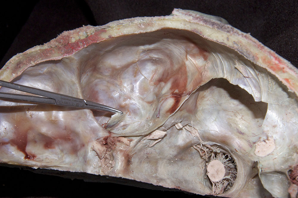
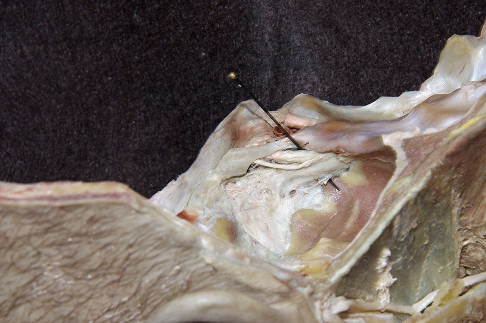
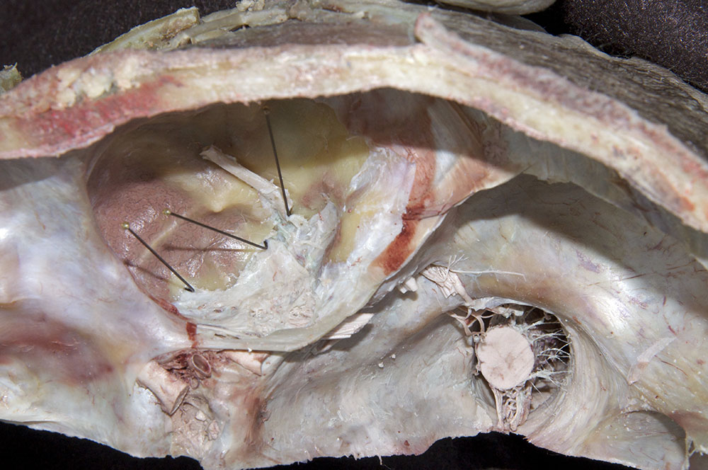
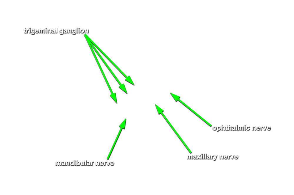
- (ON THE SIDE WITHOUT THE BRAIN) Expose the trigeminal ganglion by removing the overlying dura. Place the iris scissors between the dura and the trigeminal nerve. Open the scissors to create a slit in the dura. Wet and peel the dura from the underlying trigeminal ganglion and nerve branches. Cut the dura as needed to facilitate the peeling process. (G 7.30A;N 105;Gl 37.9) Do not remove the dura (including the tentorium cerebelli) from the posterior cranial fossa.
- (ON THE SIDE WITHOUT THE BRAIN) Identify the trigeminal ganglion, mandibular nerve at foramen ovale, maxillary nerve at foramen rotundum and ophthalmic nerve in the lateral wall of the cavernous sinus.
-
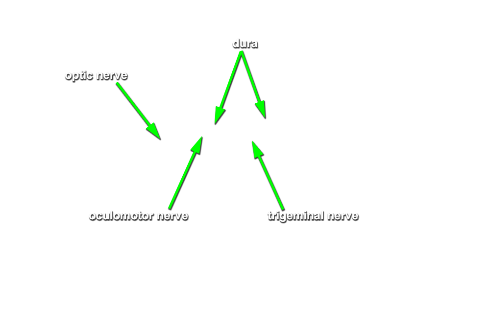
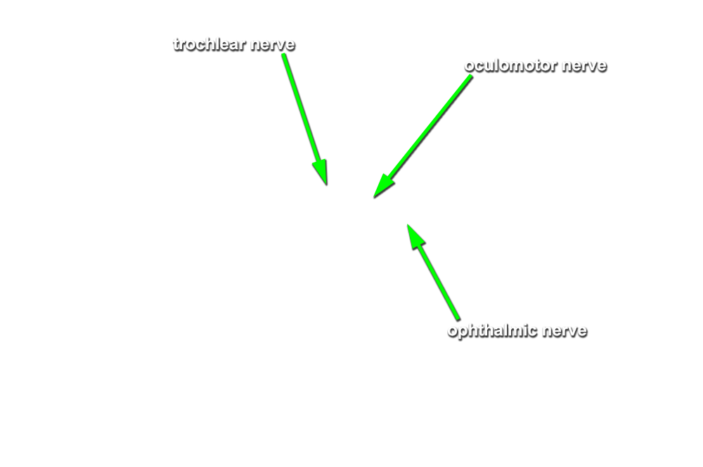
(ON THE SIDE WITHOUT THE BRAIN) Identify the
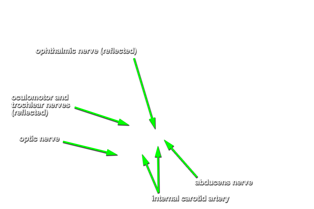
oculomotor nerve in the lateral wall of the cavernous sinus adjacent to the ophthalmic nerve. Attempt to identify the trochlear nerve. Reflect the oculomotor nerve to identify the
internal carotid artery in the cavernous sinus. Attempt to identify the
abducens nerve directly lateral to the internal carotid artery.
Important Relationships at the level of the cavernous sinus
- The internal carotid artery is positioned lateral to the pituitary.
- The abducens nerve passes directly lateral to the internal carotid artery.
- The oculomotor, ophthalmic, and trochlear nerves all pass lateral to the internal carotid artery.
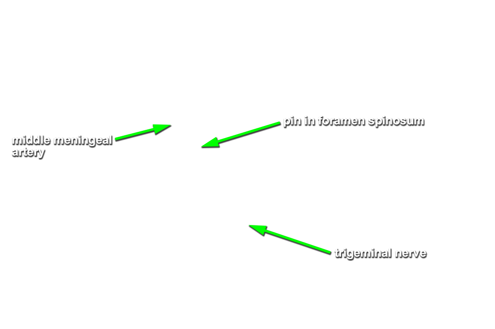
- (ON THE SIDE WITHOUT THE BRAIN) Identify the middle meningeal artery emerging from foramen spinosum. If you like challenges, then attempt to identify the greater and lesser petrosal nerves.