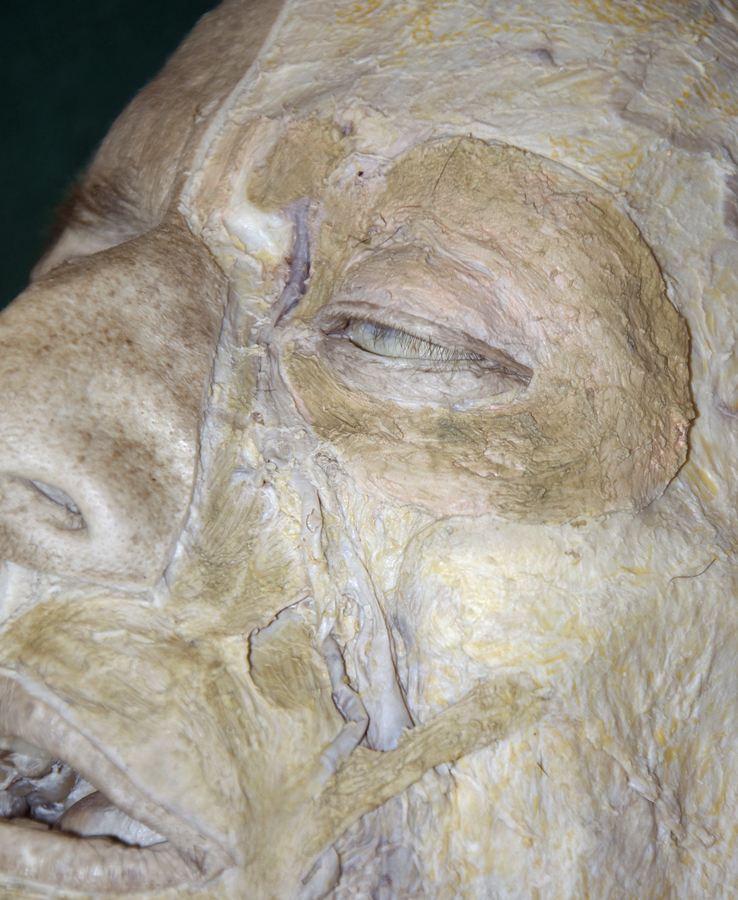
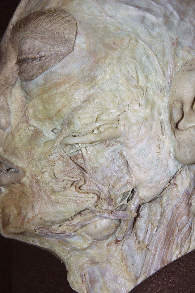
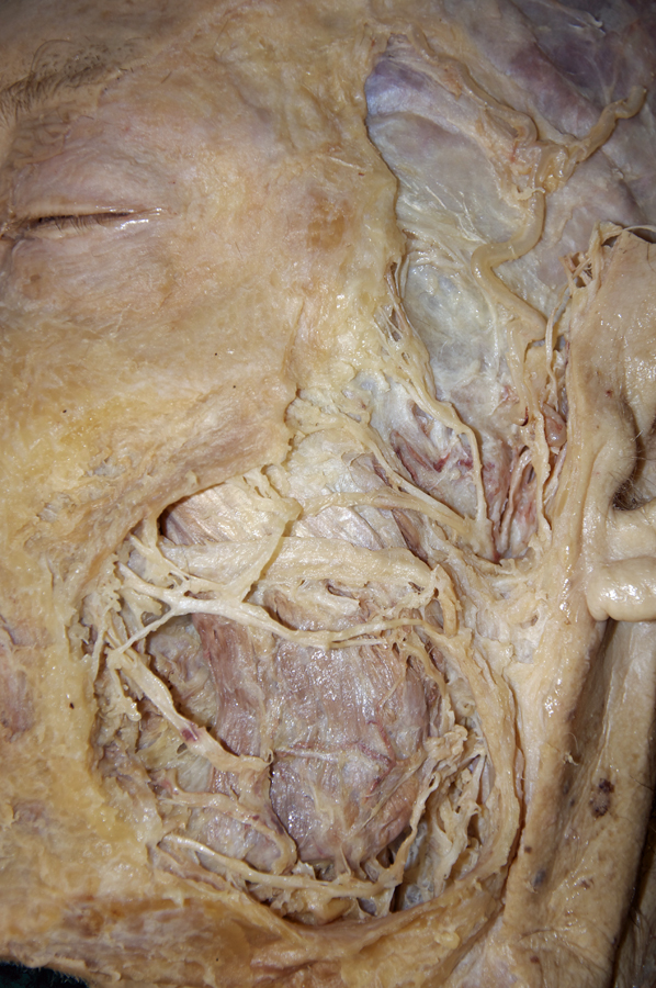
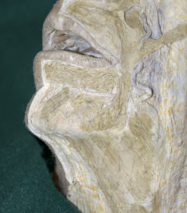
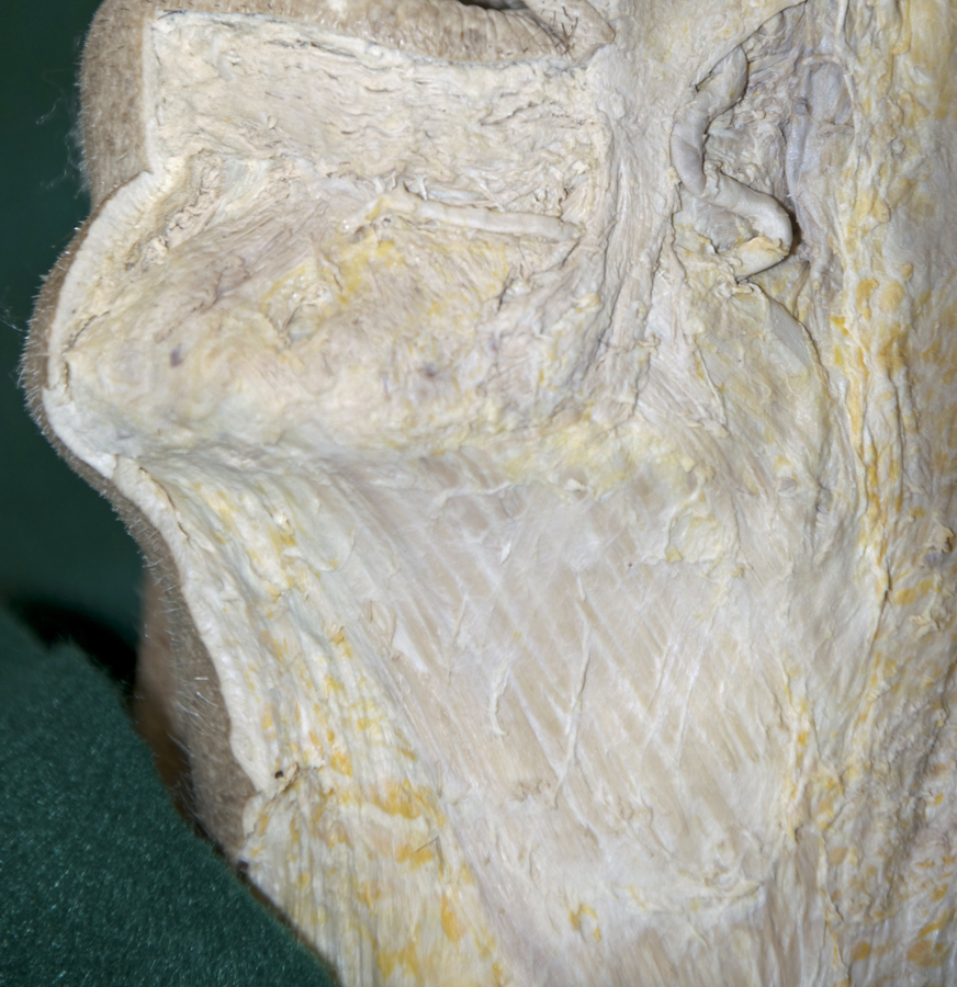
(ON THE LEFT SIDE ONLY) Identify and clean the muscles of facial expression, the parotid gland and duct, and the branches of the facial nerve. In Step 3, you will identify and clean arteries, veins and cutaneous branches of the trigeminal nerve on the right side of the face (refer to the interactive photo above for illustrations of the muscles of facial expression and the facial nerve).
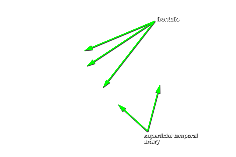
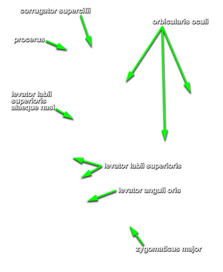
- Identify and clean the frontalis and orbicularis oculi muscles. (G 7.12;N 25;G 38.1) Attempt to distinguish the orbital and palpebral parts of the orbicularis oculi (N 25;Gl 38.1).
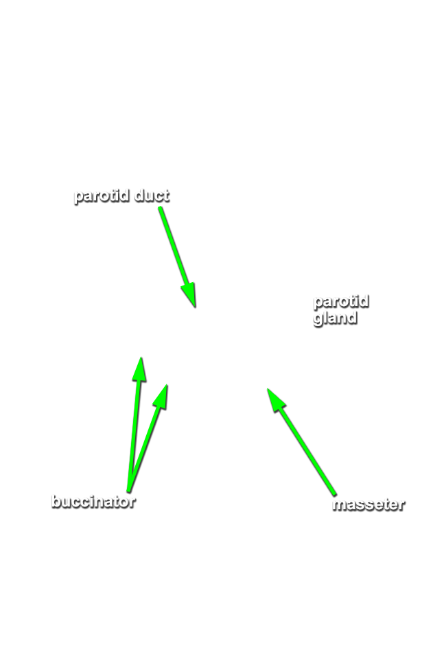
- Identify and clean the
parotid gland and
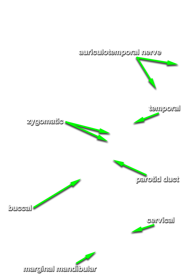
duct. (G 7.12;N 24;Gl 40.21) Note (in the illustration) the orientation of the facial nerve branches adjacent to the parotid duct. Use the scissors technique to expose one of the facial nerve branches adjacent to the parotid duct. Trace this branch back the parotid gland. Blunt dissect through the gland (remove the gland as necessary) and expose all the facial nerve branches traversing the parotid gland. Identify the
temporal,
zygomatic,
buccal,
mandibular and
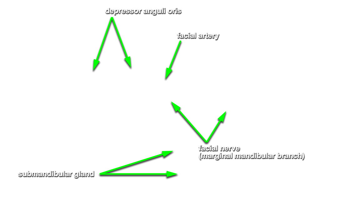
cervical branches of the facial nerve. (G 7.13;N 24;Gl 40.21) Trace (expose and clean using the scissor technique) the facial nerve branches distally until you approach the muscles of facial expression. The marginal mandibular and cervical branches are often difficult to locate as they emerge from the parotid gland. The marginal mandibular branch can be located where it passes directly superficial to the facial artery and vein as these vessels pass lateral to the body of the mandible. The cervical branch can be located on the deep surface of the platysma muscle near the angle of the mandible.
Important Relationships
- The parotid duct passes lateral (superficial) and anterior to the masseter muscle.
- The parotid gland is positioned posterior and lateral (superficial) to the masseter muscle.
- The branches of the facial nerve pass lateral (superficial) to the masseter muscle.
- Expose and identify the muscles of facial expression associated with the mouth. Start cleaning at the buccal angle (corner of the mouth) and identify the
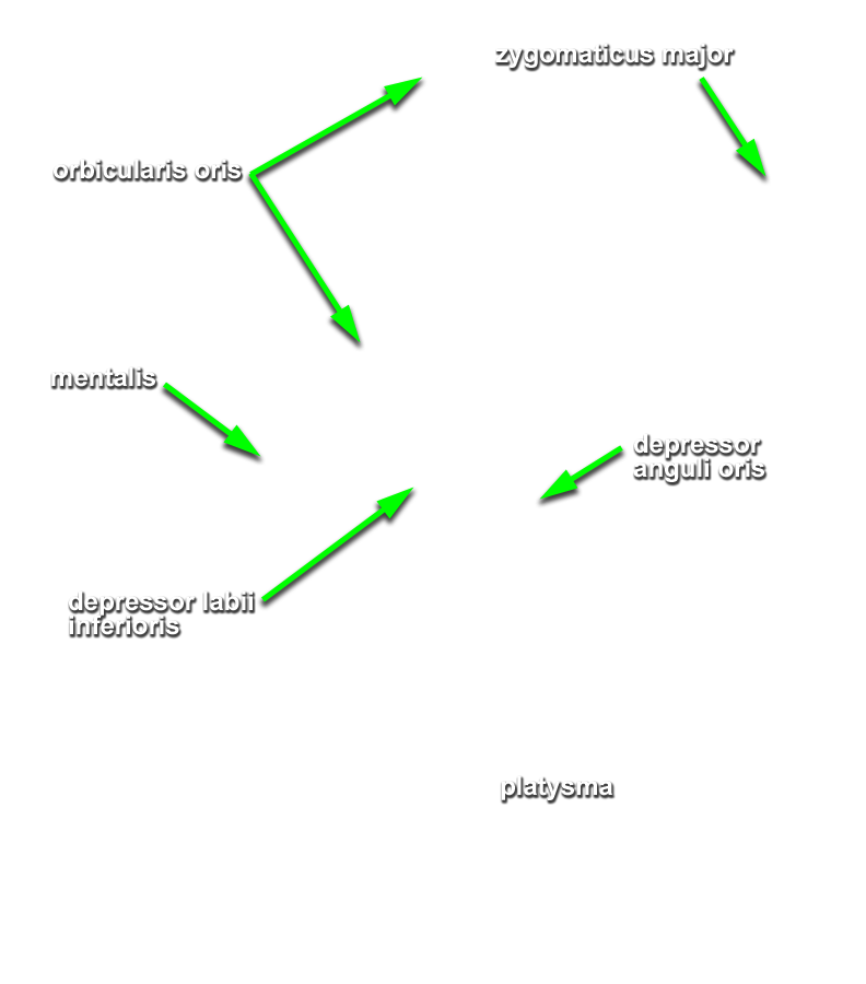
orbicularis oris,
levator anguli oris,
zygomaticus major and
depressor anguli oris muscles. (G 7.12;N 25;Gl 38.1) Continue cleaning medial to the levator anguli oris and expose and identify the
levator labii superioris muscle. Attempt to identify the
alaeque nasi part of this muscle. (G 7.12;N 25;Gl 38.1) Clean medial to the depressor anguli oris and attempt to identify the
depressor labii inferioris and
mentalis muscles. Clean lateral to the angle of the mouth (remove the buccal fatpad) to expose and identify the
buccinator muscle. (G 7.16;N 48;Gl 40.21)
If you cannot locate the buccinator muscle, then place the handle of your forceps in the oral cavity and push out on the cheek to bring the buccinator muscle to a more superficial position. - Identify and clean the platysma muscle where it attaches to the mandible. (G 7.13;N 25;Gl 38.1) You do not need to clean the platysma muscle overlying the anterior-lateral neck at this time.
- Return to the facial nerve branches. If times permits, attempt to trace the facial nerve branches to the exposed and cleaned muscles of facial expression. Don't panic if the artery, vein and sensory nerve dissection on the opposite side of the face is completed and you have not finished cleaning the muscles and facial nerve branches. The infratemporal fossa dissection is only performed on the opposite (right) side of the head (artery, vein and cutaneous nerve side). You can finish cleaning the muscles and facial nerve branches during the infratemporal fossa dissection.