(ON THE RIGHT SIDE ONLY) Identify and clean arteries, veins and cutaneous branches of the trigeminal nerve. You will destroy many of the muscles of facial expression as you clean the arteries, veins and nerves (refer to the interactive photo above for illustrations of the arteries of the face and cutaneous, trigeminal nerve distribution).
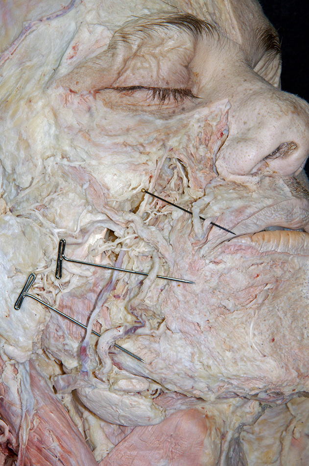
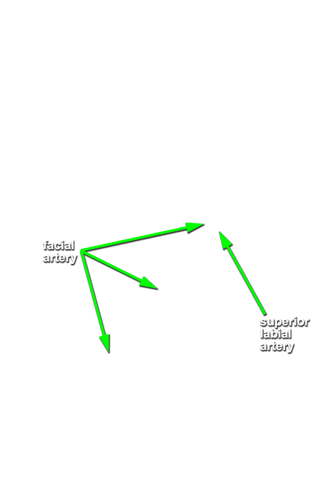
- Blunt dissect immediately lateral to the angle of the mouth to expose and identify the
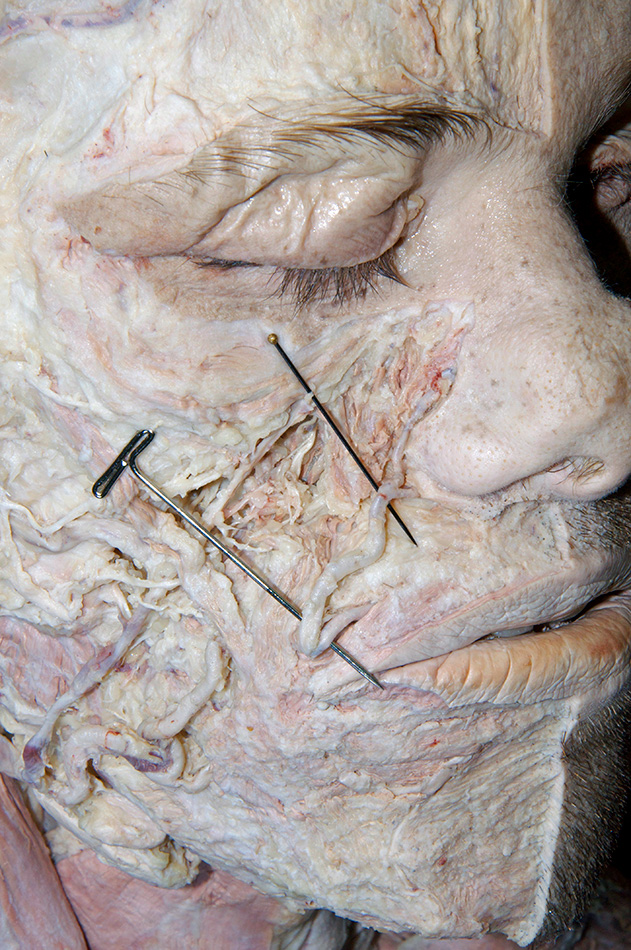
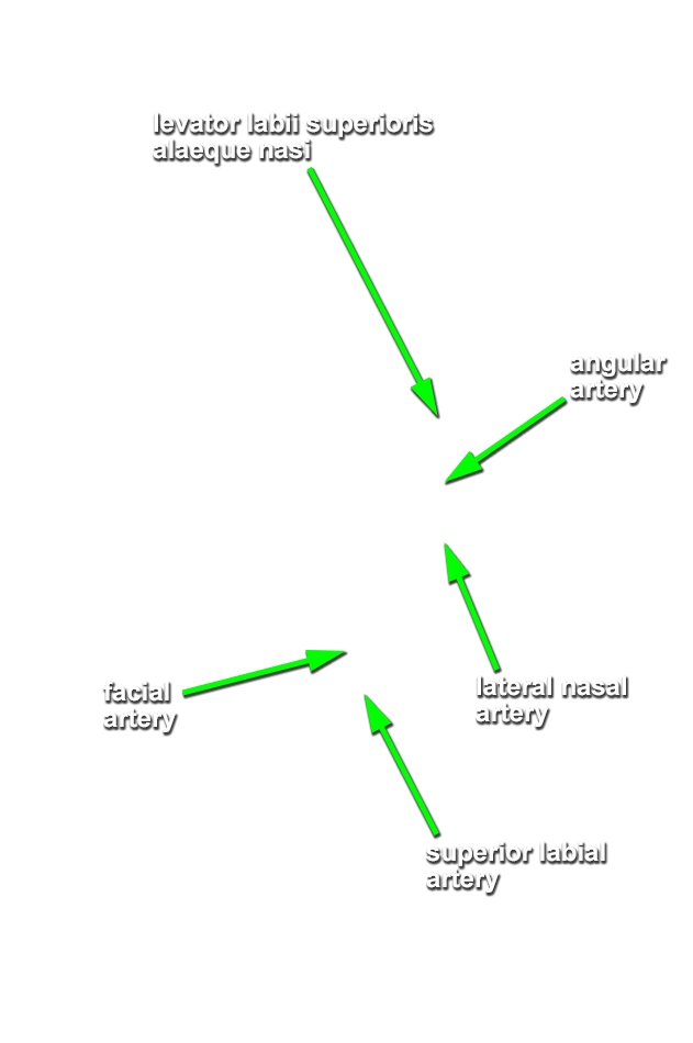
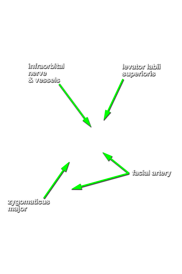
facial artery. (G 7.12;N 3;Gl 40.4) The facial artery typically has a superficial loop at this location. Trace (blunt dissect) the facial artery in the superior direction. Identify the superior labial artery and attempt to identify the
lateral nasal and
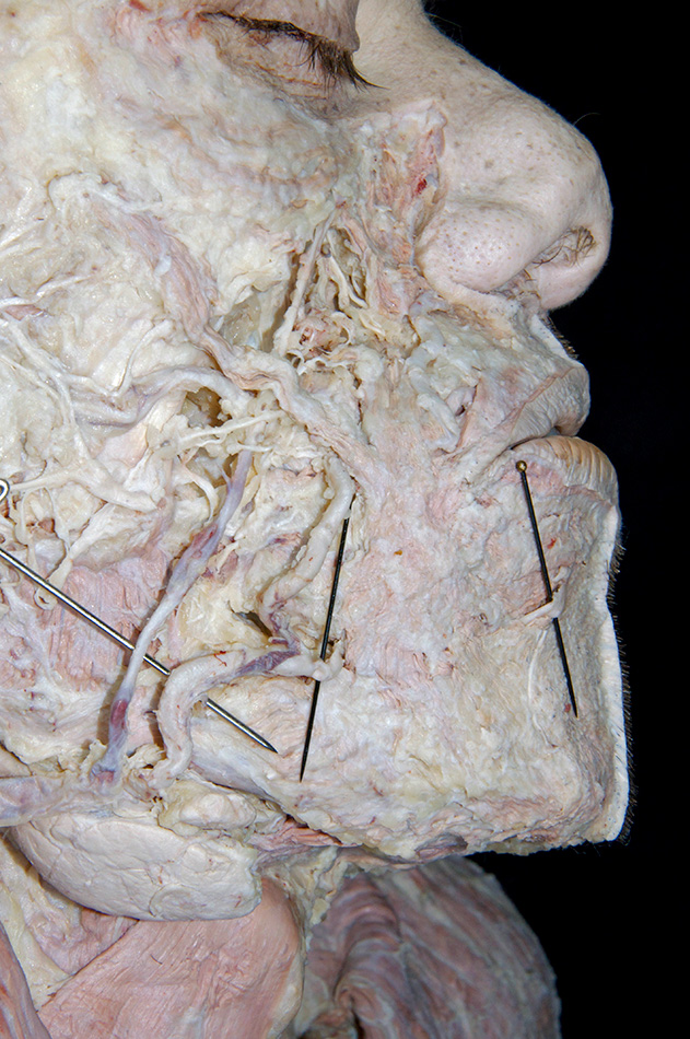
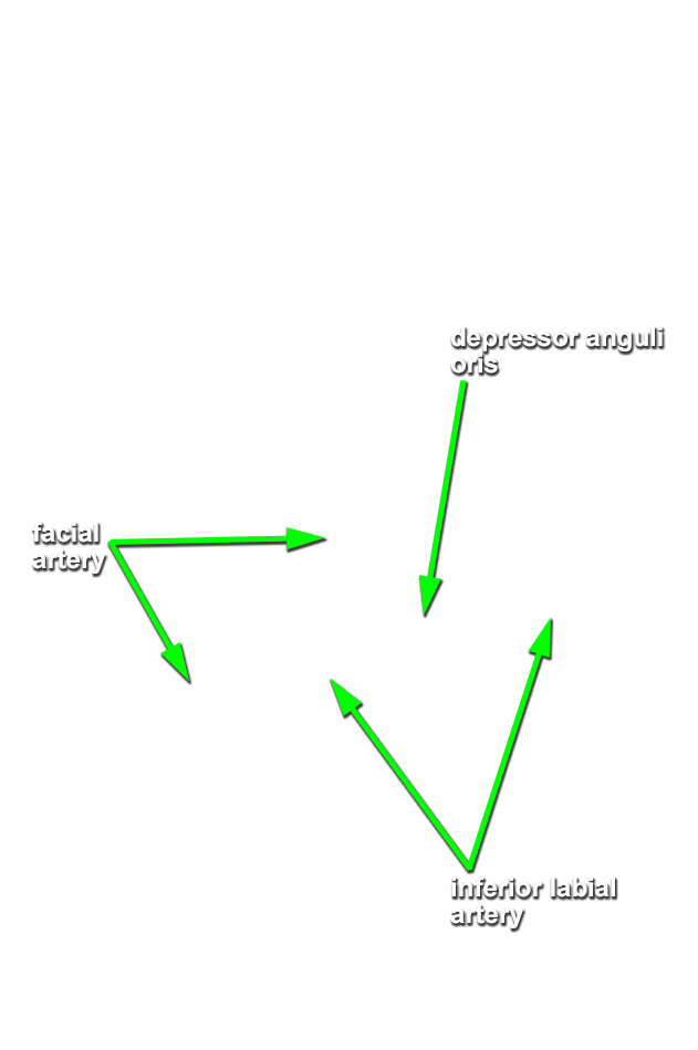
angular (the terminal branch) arteries. (G Table 7.18;N 3;Gl 40.4) Trace the facial artery in the inferior direction until it crosses the mandible. Identify the inferior labial artery.
Important Relationship
- The facial artery passes lateral (superficial) to the mandible (body).
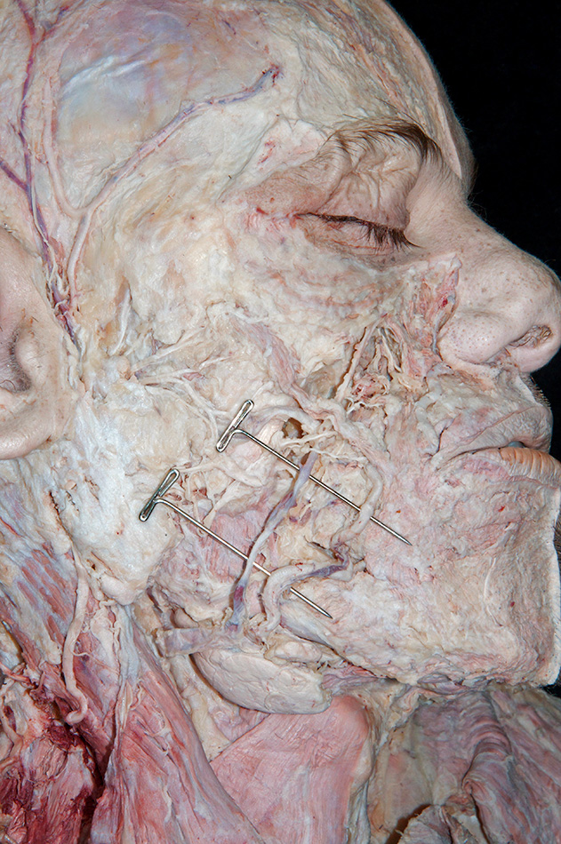
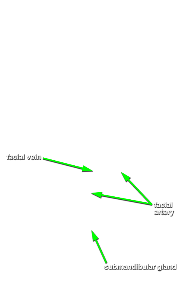
- Blunt dissect immediately posterior to the facial artery where it passes lateral to the mandible to expose and identify the
facial vein. (G 7.12;N 3;Gl 40.9B) Trace the facial vein in the superior direction towards the lateral aspect of the nose.
Important Relationship
- On the face, the facial vein is positioned posterior to the facial artery.
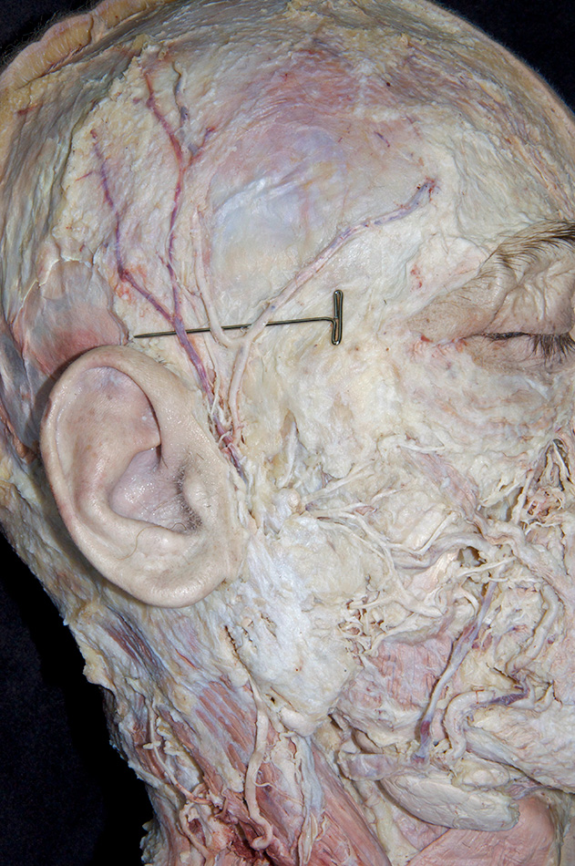
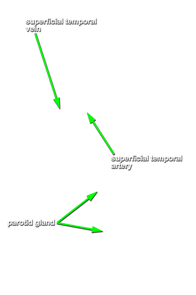
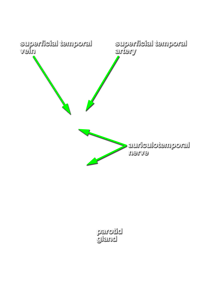
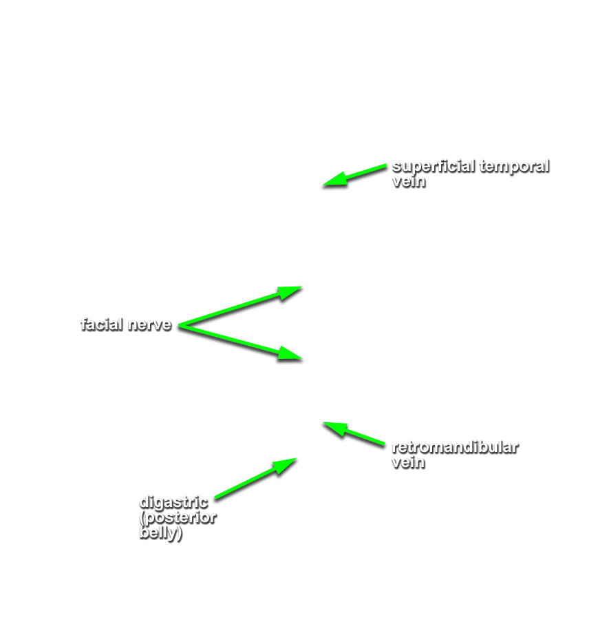
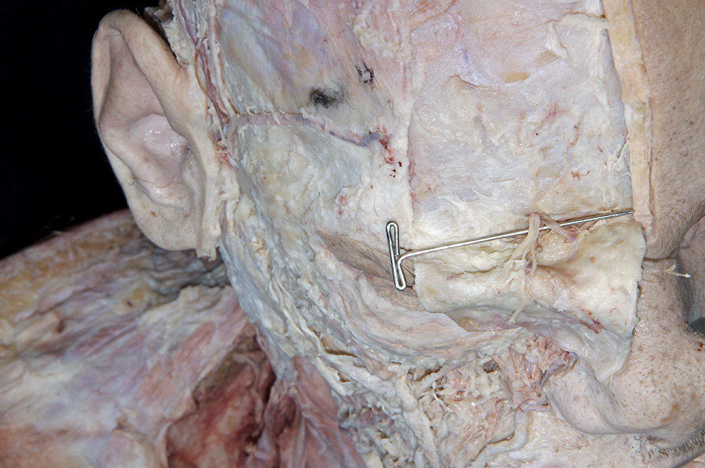
- Expose and identify the superficial temporal artery and superficial temporal vein anterior to the ear and superior to the parotid gland. (G 7.12;N 3;Gl 40.9B) Carefully blunt dissect adjacent and parallel to the superficial temporal artery to expose and identify the auriculotemporal nerve. (G 7.12;N 2;Gl 40.21) You will need this nerve when you dissect the infratemporal fossa.
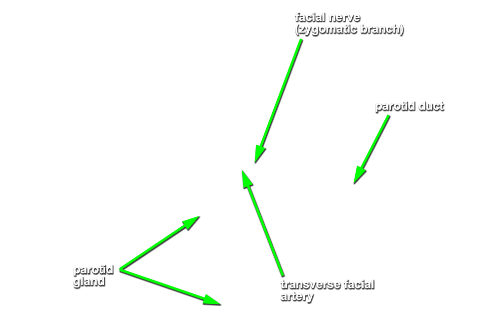
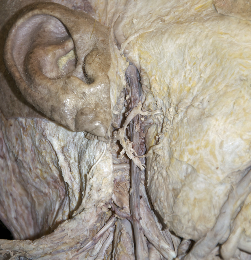
- Expose and identify the parotid duct. Attempt to identify the transverse facial artery immediately superior to the parotid duct. (G 7.12;N 3;Gl 40.21)
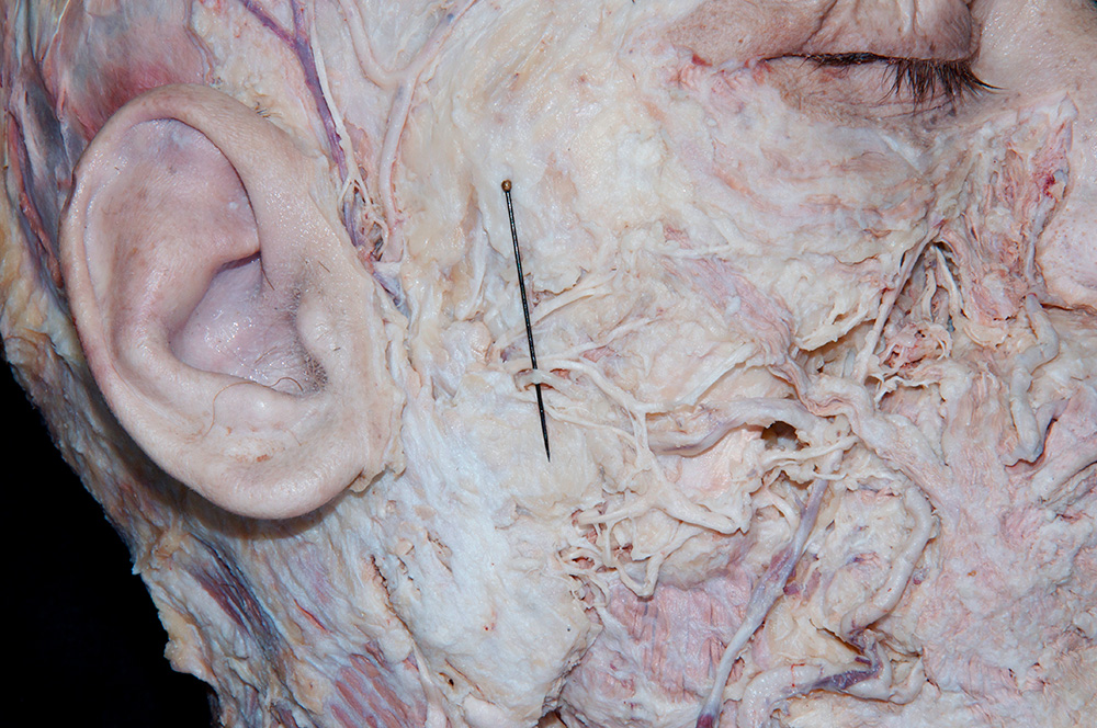
- Return to the superficial temporal artery and vein. Fully expose these structures by blunt dissecting through and removing the parotid gland. Preserve the trunks of the facial nerve when you encounter these structures.
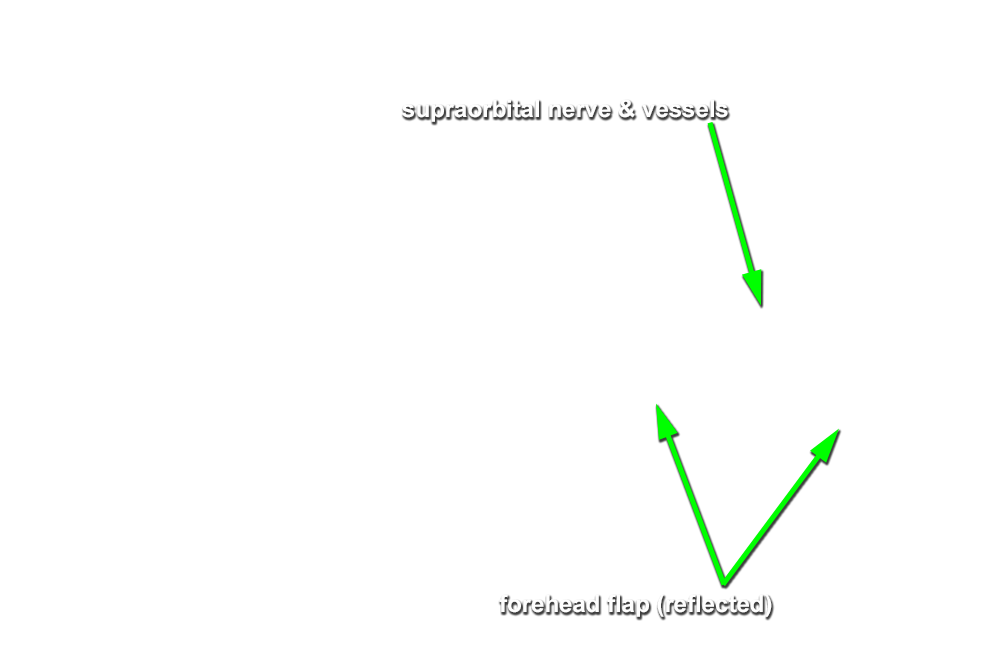
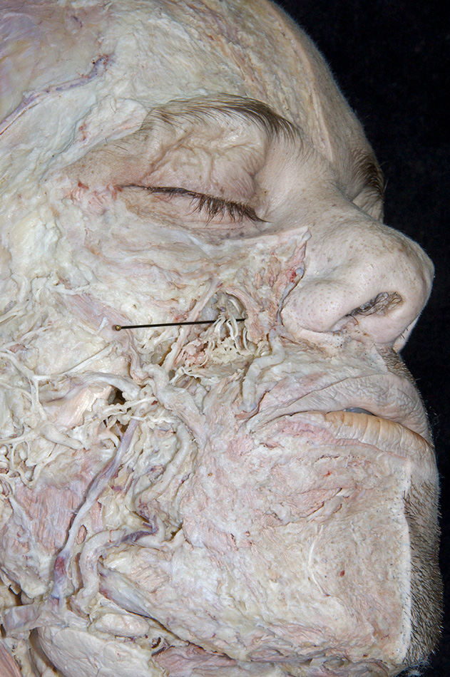
- Use a scalpel blade and make a transverse incision through the frontalis muscle and fascia approximately 5 cm superior to the supraorbital margin. Extend the incision in the lateral direction as far as the lateral margin of the orbit. Make two vertical incisions (the first along the midline and the second parallel with the lateral margin of the orbit) to create a flap of muscle and fascia. Blunt dissect the flap away from the underlying frontal bone (in the inferior direction) until you reach the supraorbital margin. Expose and identify the supraorbital nerve and artery. (G 7.16;N 3 and 2;Gl 41.10) If time permits, attempt to expose and identify the supratrochlear, infratrochlear, external nasal, zygomaticofacial and zygomaticotemporal nerves.
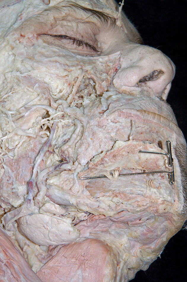
- Clean deep to the levator labii superioris and orbicularis oculi muscles to expose and identify the infraorbital nerve and artery. (G 7.16;N 3 and 2;Gl 41.10) The nerve and artery can typically be located approximately 1 cm inferior to the infraorbital margin in line with the first molar. Trace the nerve and artery in the superior direction to the infraorbital foramen.
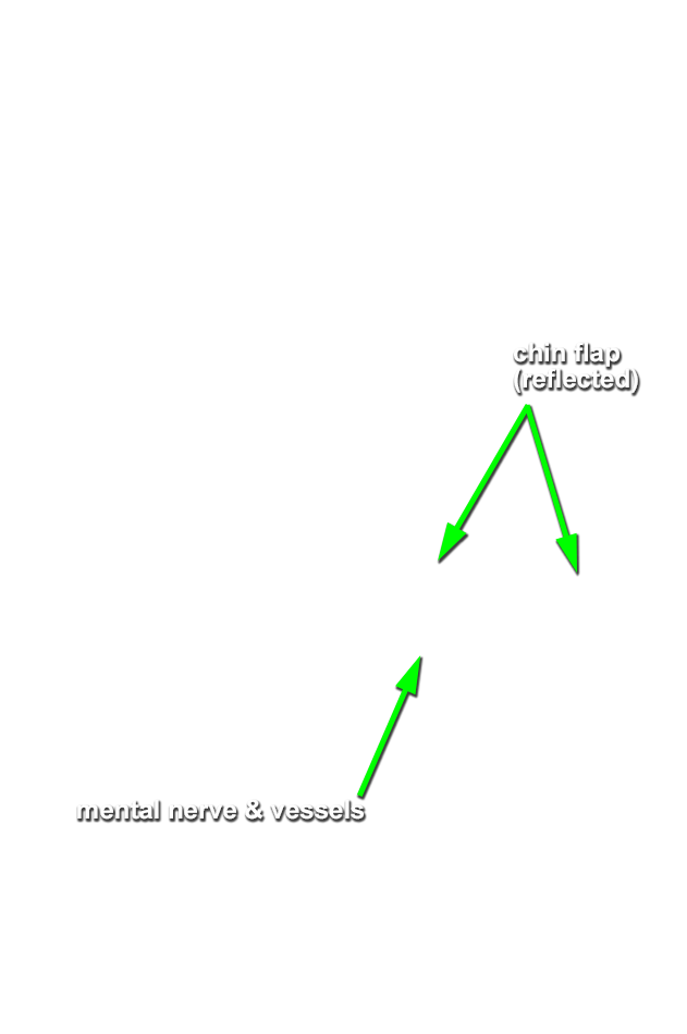
- Make a midline incision through the fascia and muscle covering the lower half of the chin to the level of the mental protuberance. Carry the incision in the posterior-lateral direction along the margin of the mandible as far as the second molar. Blunt dissect the tissue covering the chin away from the bone in the superior-lateral direction to expose and identify the mental nerve and artery. (G 7.16;N 3 and 2;Gl 40.20) The nerve and artery typically emerge from the mental foramen in line with the first premolar.