Only do this infratemporal fossa dissection on the right side of the head (the artery, vein and sensory nerve dissection).
STOP
BEFORE PROCEEDING WITH THIS DISSECTION
READ STEPS 1, 2 AND 3 AND CAREFULLY STUDY THE PHOTOGRAPHS AND ANIMATION.
YOU WILL BE MAKING FIVE CUTS:
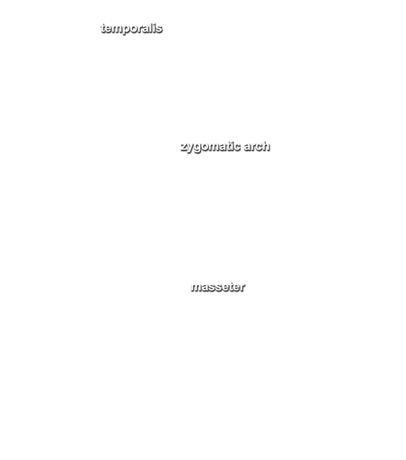
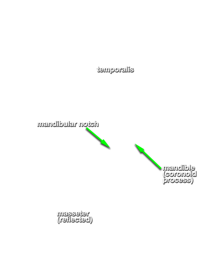
YOU WILL REFLECT THE ZYGOMATIC ARCH AND ATTACHED MASSETER MUSCLE IN THE INFERIOR DIRECTION TOWARDS THE ANGLE OF THE MANDIBLE. YOU WILL REFLECT THE CORONOID PROCESS AND ATTACHED TEMPORALIS MUSCLE IN THE SUPERIOR DIRECTION TOWARD THE TEMPORAL FOSSA. YOU WILL LEAVE THE HEAD OF THE MANDIBLE WITH THE TMJ (UNTIL STEP 4). YOU WILL REMOVE THE CUT PORTION OF THE RAMUS OF THE MANDIBLE BETWEEN SAW CUTS 3 AND 4 AND 5.
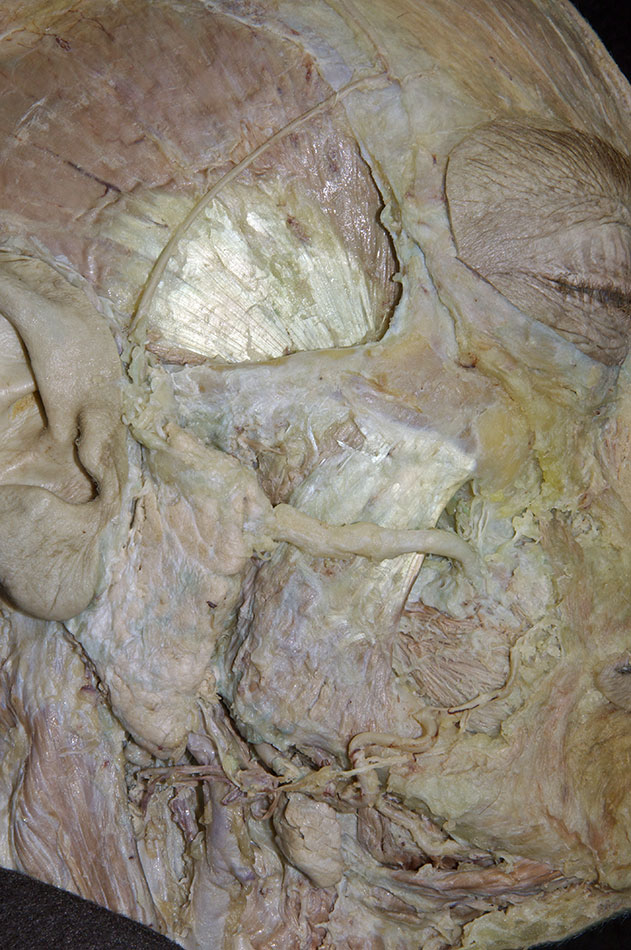
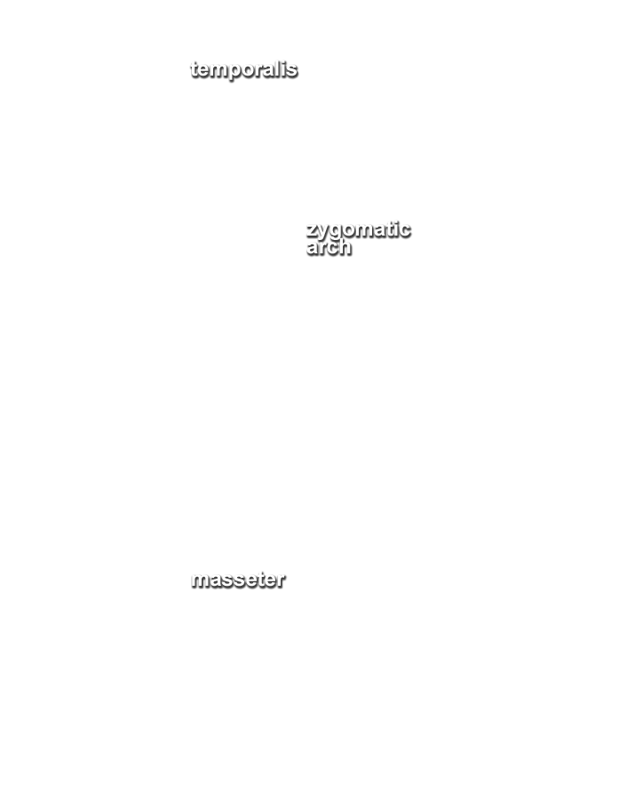
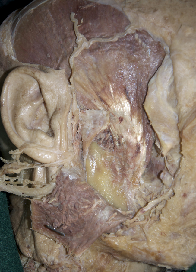
Identify, clean and reflect the masseter muscle. (G 7.12;N 48; Gl 40.21)
- (ON THE RIGHT SIDE ONLY) Clean the superficial aspect of the masseter muscle by removing the fascia and parotid gland and duct. Leave at least one branch of the facial nerve dangling near the ear as you remove the parotid gland.
- (ON THE RIGHT SIDE ONLY) Identify and clean the zygomatic arch. Cut the deep fascia and blunt dissect along the superior aspect of the zygomatic arch separating the arch from the underlying temporalis muscle.
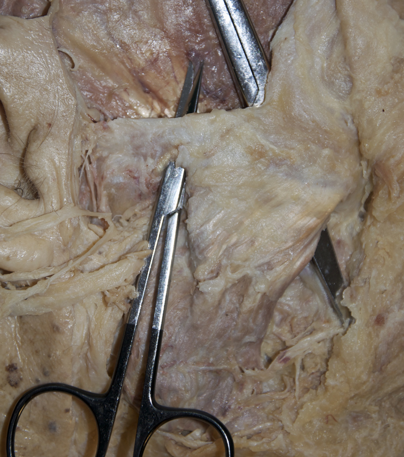
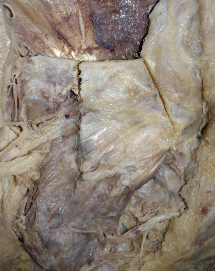
- (ON THE RIGHT SIDE ONLY) Use the electric saw to cut the zygomatic arch as far anterior as possible (cut 1). Make a second cut (cut 2) in the arch immediately anterior to the articular tubercle and eminence.
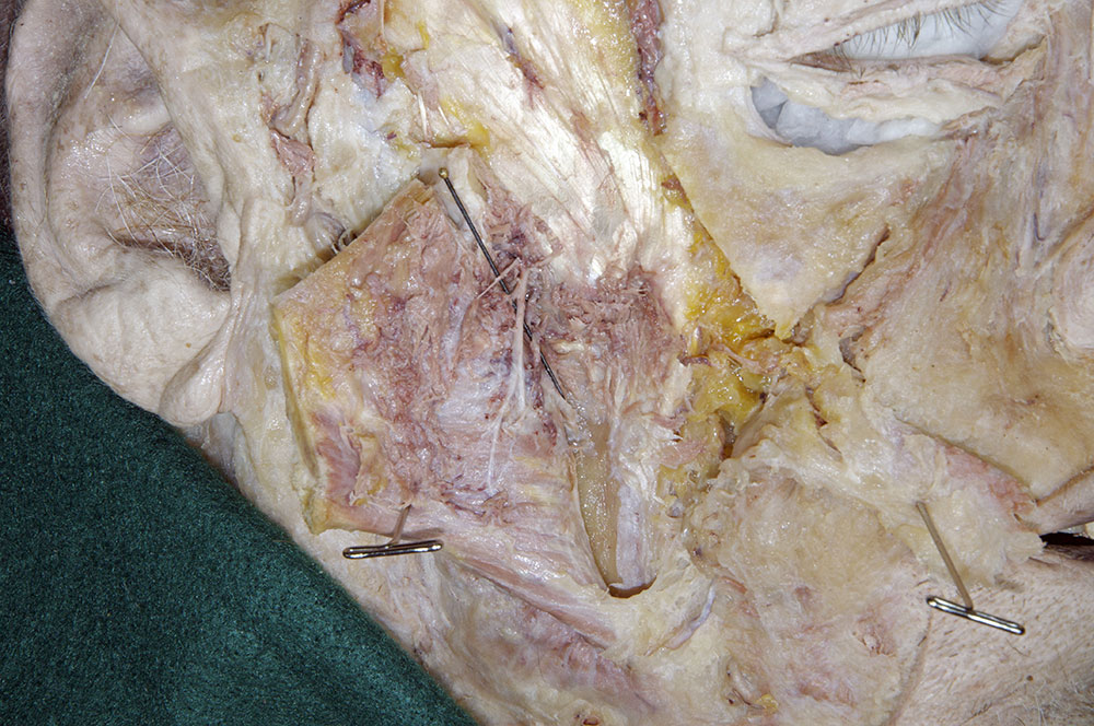
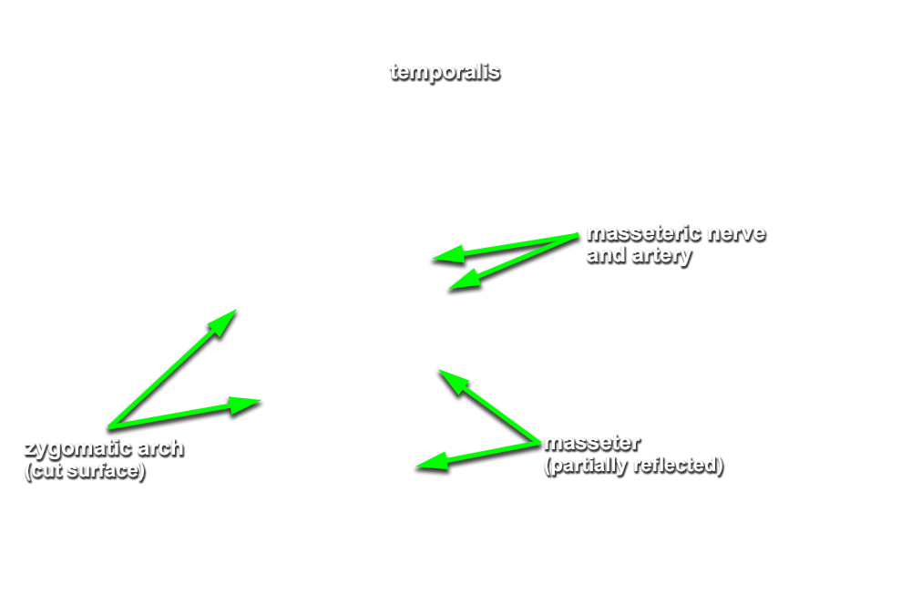
- (ON THE RIGHT SIDE ONLY) Reflect the zygomatic arch and masseter muscle in the inferior direction towards the angle of the mandible. (G 7.49;N 49; Gl 38.2) Leave the masseter attached to the angle of the mandible. As you reflect the masseter muscle, attempt to identify the masseteric nerve and artery near the mandibular notch.
- The masseter muscle is positioned lateral (superficial) to the mandible (ramus) and inferior to the zygomatic arch.