Identify and clean the structures of the carotid triangle.
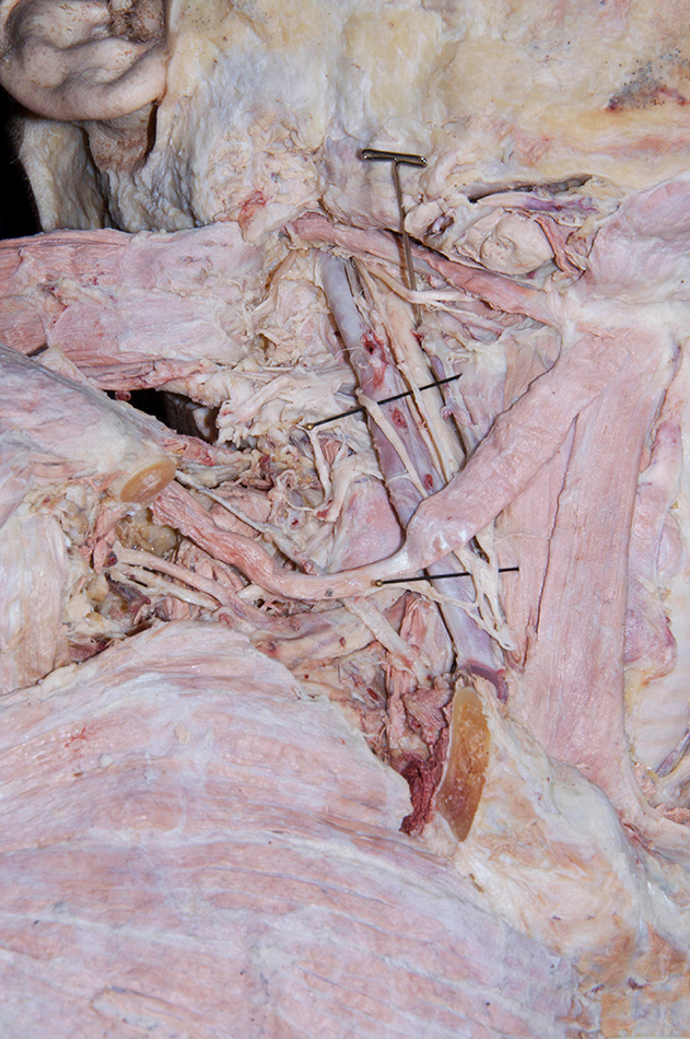
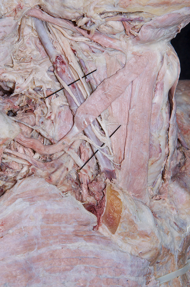
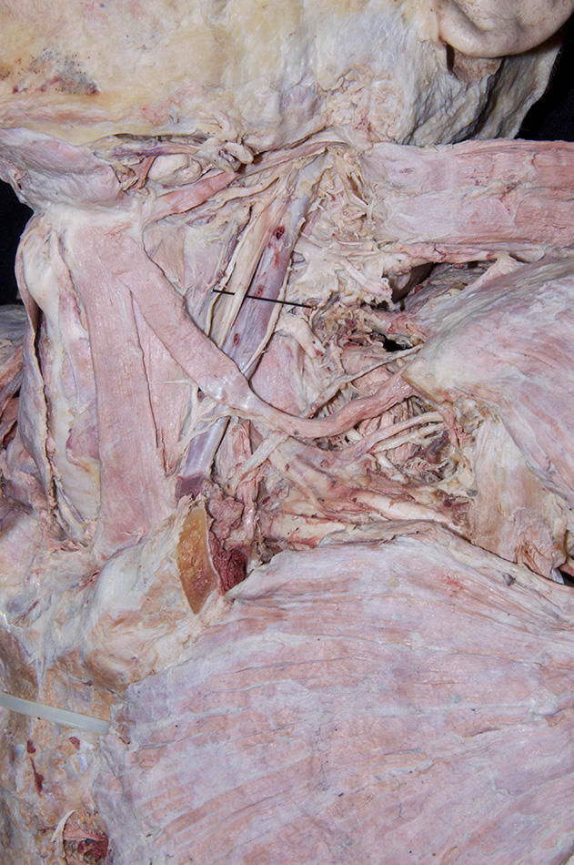
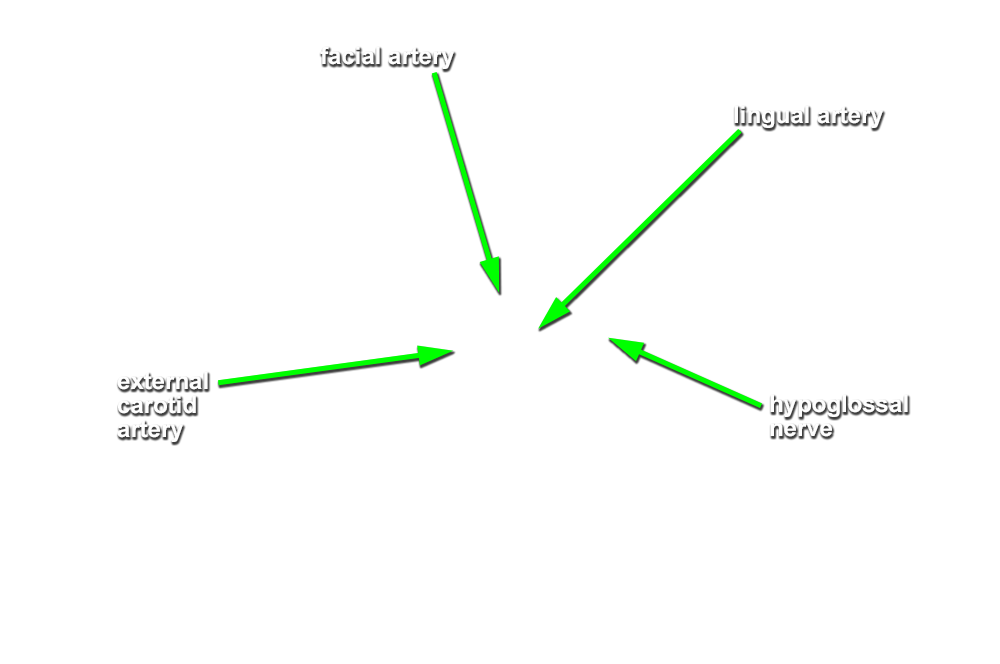
- (ON BOTH SIDES) Identify the carotid sheath and the associated ansa cervicalis. (G 8.14;N 32;Gl 44.17C) Carefully trace the branches of the ansa cervicalis to the infrahyoid muscles. The branches on the right side may have been torn when the infrahyoid muscles were reflected in the previous step.
- The ansa cervicalis (inferior root) passes lateral (superficial) to the internal jugular vein.
- The ansa cervicalis (superior root) is positioned anterior to the internal jugular vein.
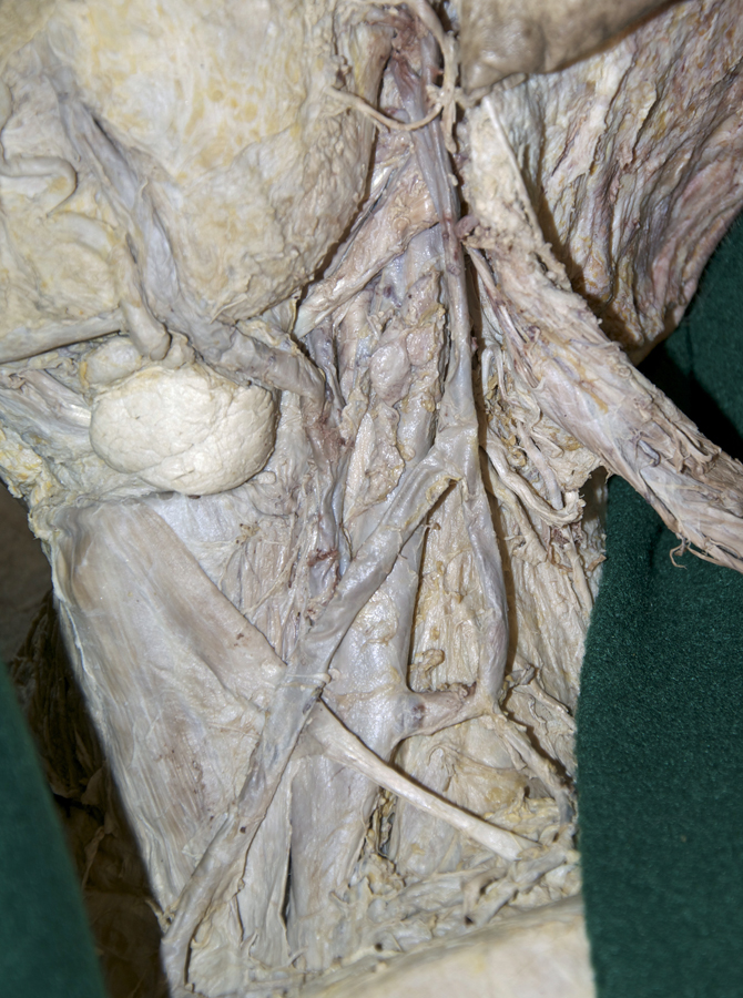
- (ON BOTH SIDES) Blunt dissect through the carotid sheath, remove any deep cervical lymph nodes, and identify the common carotid artery, internal jugular vein, and vagus nerve. (G 8.18;N 26;Gl 45.32)
- The vagus nerve is positioned posterior-medial to the internal jugular vein and posterior-lateral to the common carotid artery.
- The internal jugular vein is positioned lateral to the carotid artery.
- The external carotid artery is positioned anterior to the internal carotid artery.
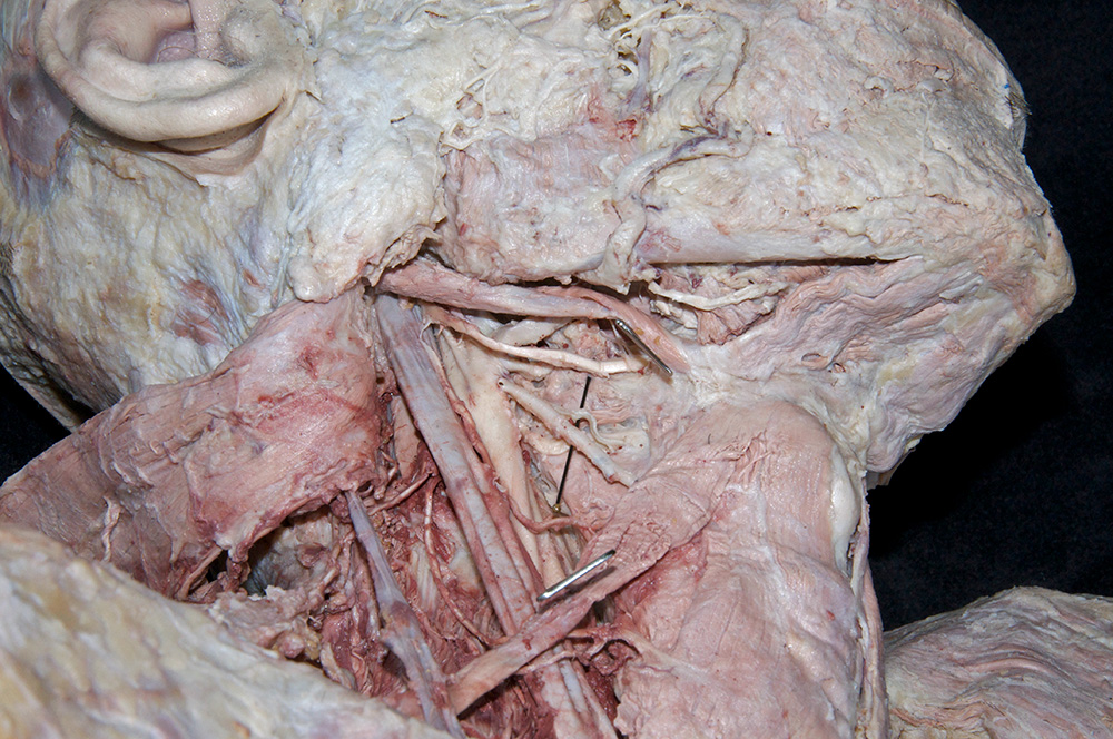
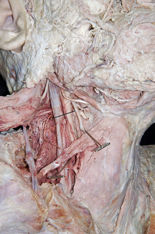
- (ON BOTH SIDES) Trace (clean) the common carotid artery in the superior direction until it bifurcates (near the level of the hyoid bone) into the internal and external carotid arteries.
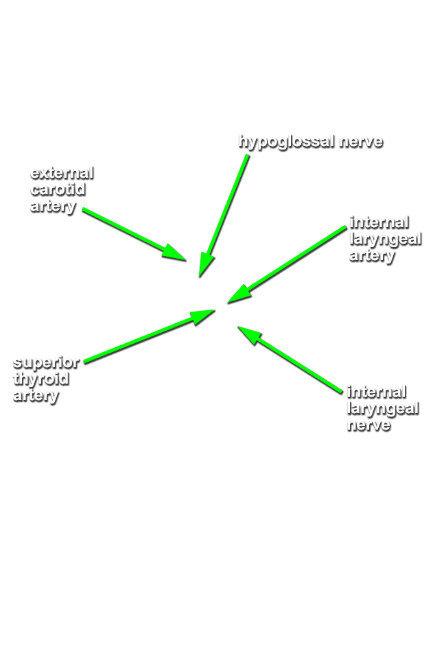
- (ON THE RIGHT SIDE ONLY) Identify and clean the superior thyroid artery. (G 8.18;N 34;Gl 45.32) Trace this vessel in the inferior direction to the thyroid gland.
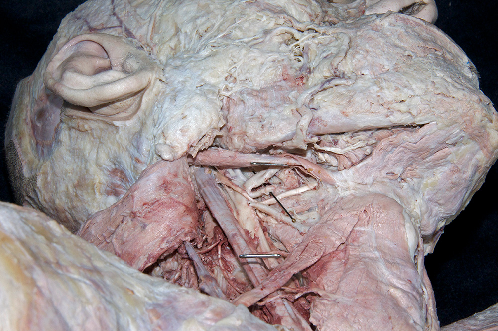
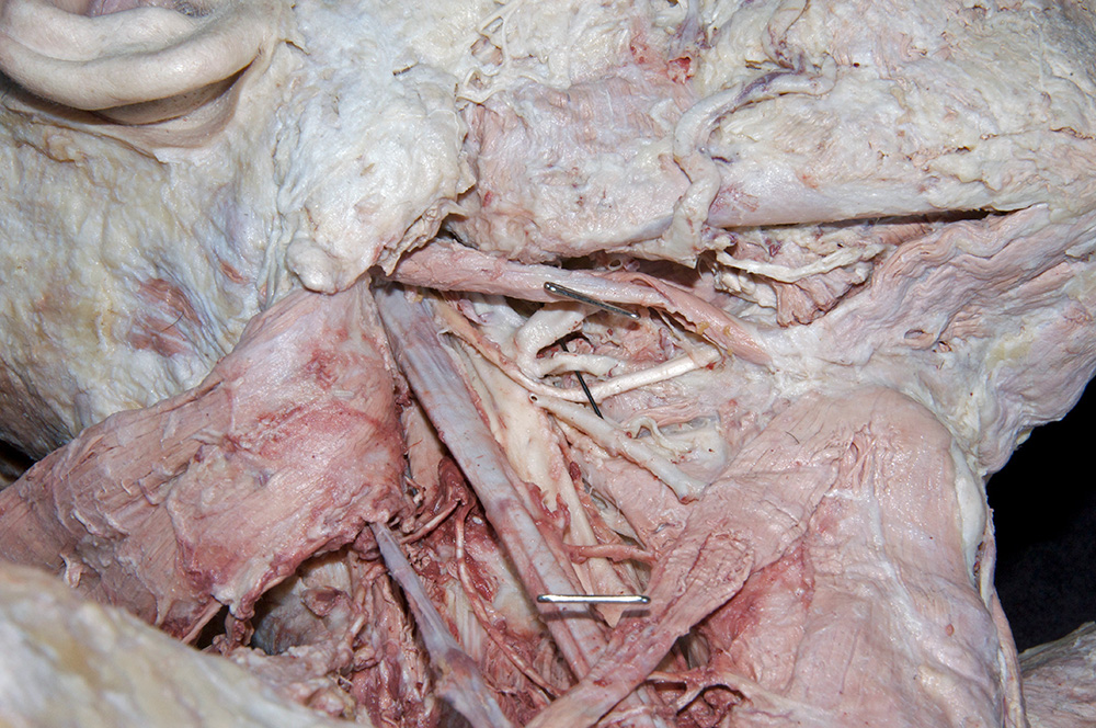
- (ON THE RIGHT SIDE ONLY) Identify and clean the facial and lingual arteries. (G 8.15;N 34;Gl 45.33) The lingual and facial arteries often have a common origin from the external carotid artery. Trace the facial artery from its origin to the face. Attempt to identify its submental branch. Trace the facial vein superficial to the mandible where it eventually drains into the common facial vein. The common facial vein is typically a tributary of the internal jugular vein. Do not trace the lingual artery to the tongue at this time.
- The facial artery passes medial (deep) to the stylohyoid muscle and the intermediate tendon of the digastric muscle.
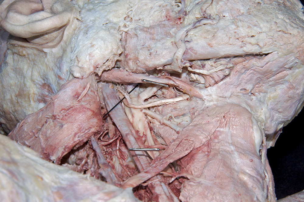
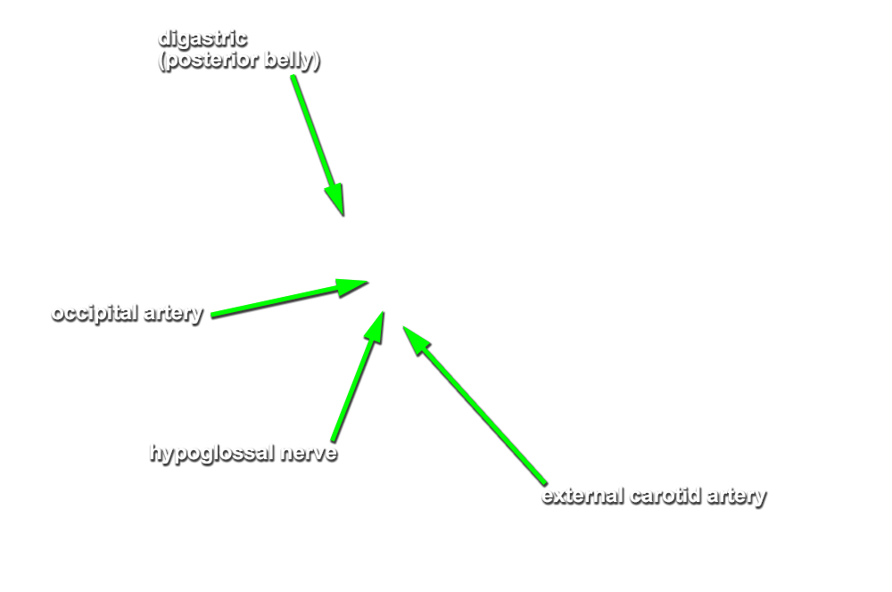
- (ON THE RIGHT SIDE ONLY) Identify the hypoglossal nerve and the occipital artery. (G 8.14;N 34;Gl 40.6) The hypoglossal nerve crosses lateral to the occipital artery and is often crossed by the SCM branch of the occipital artery. Return to the ansa cervicalis. Trace its superior root to the hypoglossal nerve. Attempt to identify the thyrohyoid nerve appearing to branch from the hypoglossal nerve. Trace the hypoglossal nerve in the anterior direction until it passes deep to the mylohyoid muscle.
- The hypoglossal nerve passes medial (deep) to the stylohyoid muscle and the intermediate tendon of the digastric muscle and lateral (superficial) to the hyoglossus muscle.
- During its posterior course, the occipital artery first passes medial (deep) and then lateral (superficial) to the hypoglossal nerve.
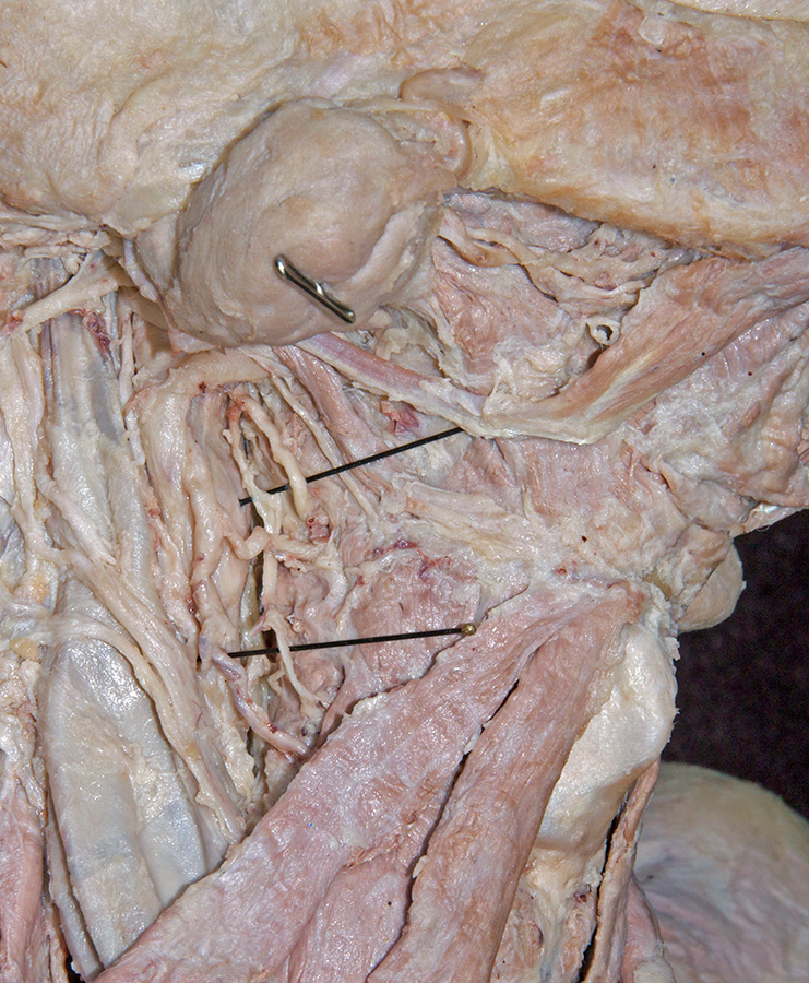
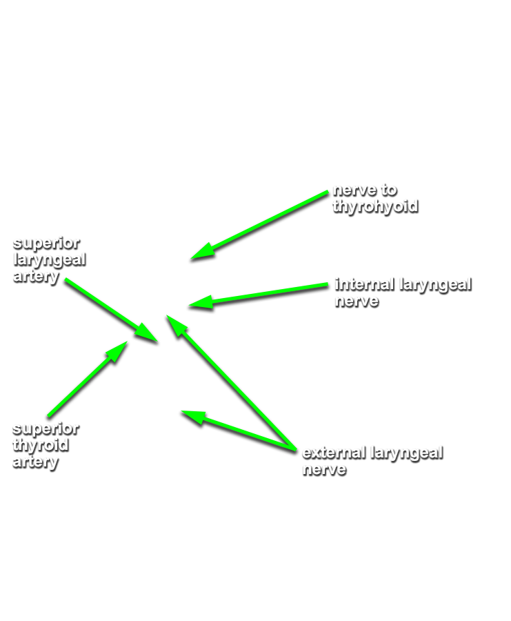
- (ON BOTH SIDES) Identify and clean the internal laryngeal branch of the superior laryngeal nerve. (G 8.20;N 76;Gl 45.28A) Locate this nerve where it pierces the thyrohyoid membrane (lateral aspect of the larynx and immediately inferior to the hyoid). Attempt to identify the superior laryngeal artery and external laryngeal nerve.
Important Relationships
Important Relationships
Important Relationships
Important Relationships