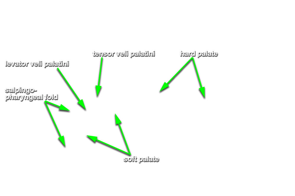
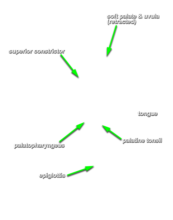
Identify the internal surface features and muscles associated with the nasal pharynx, oral pharynx and palate.
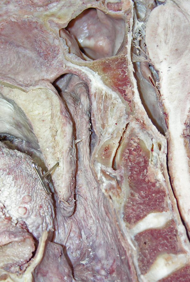
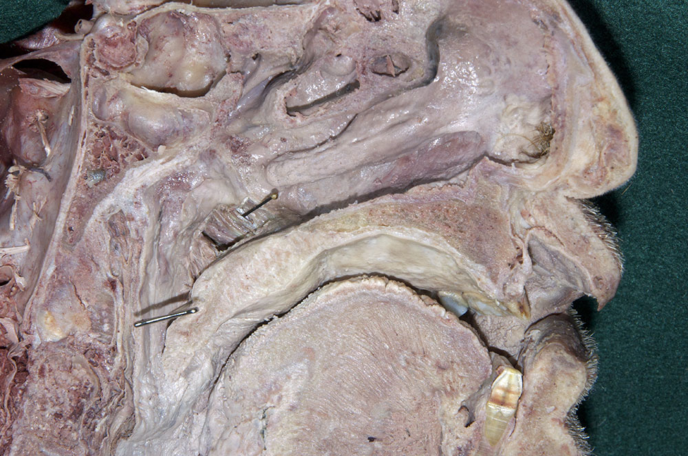
- Identify the opening and torus of the pharyngotympanic tube, ridge of the levator veli palatini, salpingopharyngeal fold and pharyngeal recess. (G 8.32A;N 68;Gl 44.23)
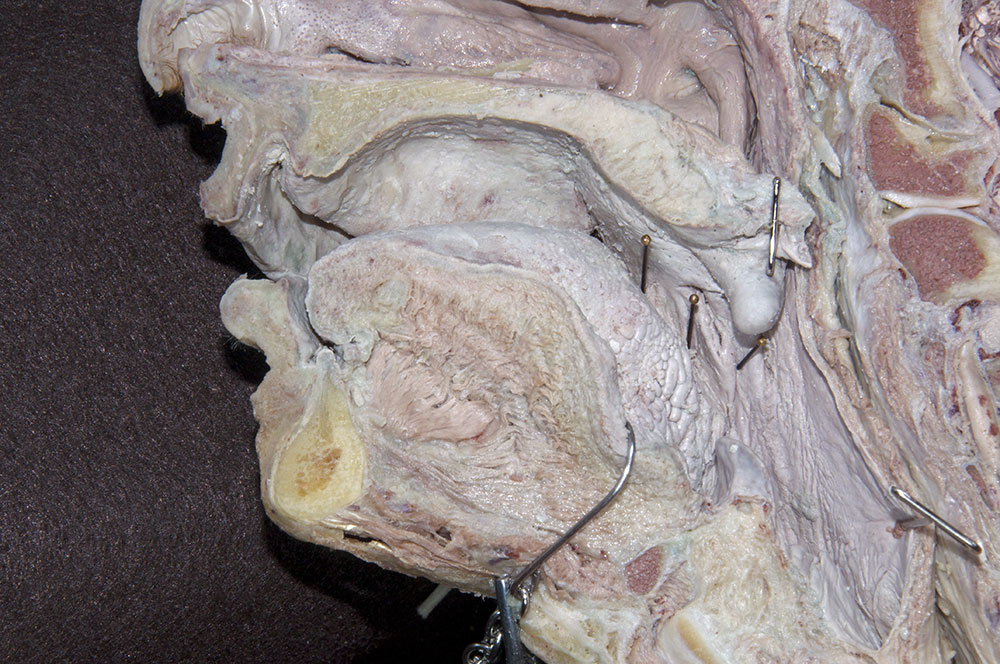
- Identify the
oropharyngeal isthmus,
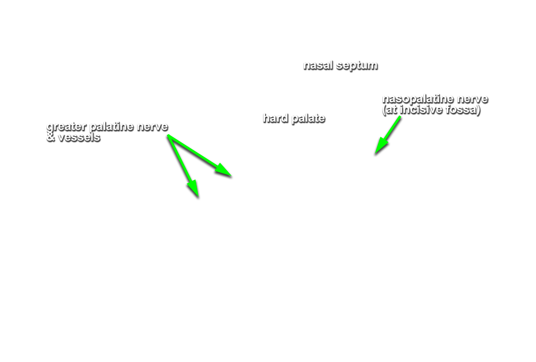
hard palate,
soft palate (in cross section but not identified) and
uvula,
palatoglossal arch (fold),
tonsilar bed (fossa) and
palatopharyngeal arch (fold). (G 8.32A;N 68;Gl 44.27)
Important Relationship
- The tonsilar bed is positioned anterior to the palatopharyngeal arch and posterior to the palatoglossal arch.
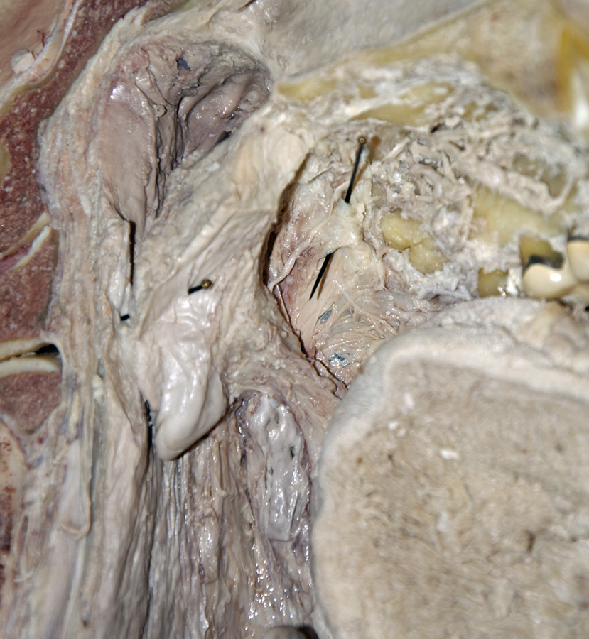
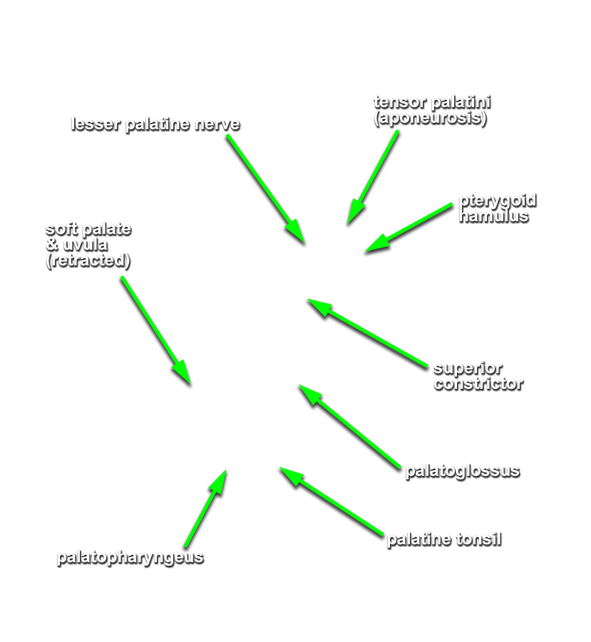
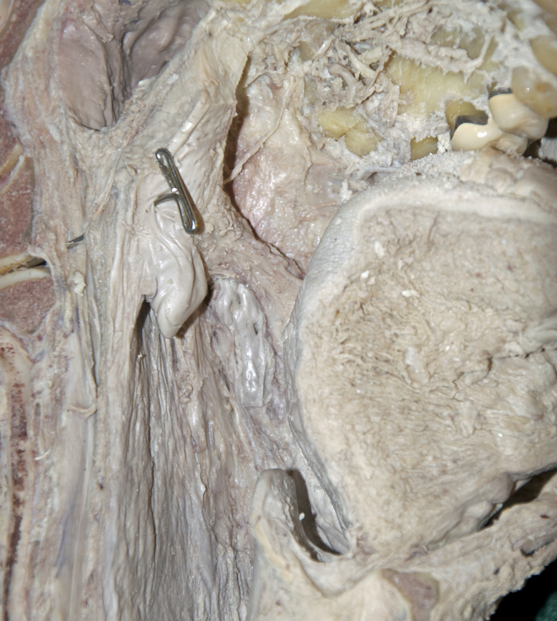
- Identify the
levator veli palatini and tensor veli palatini muscles. (G 8.32B;N 65;Gl 44.29B) The superior constrictor passes between the tensor veli palatini and the levator veli palatini muscles. Attempt to trace the tendon of the tensor veli palatini to the hamulus of the medial pterygoid plate.
Important Relationship
- The tensor veli palatini muscle is positioned anterior-lateral to the levator veli palatini muscle.
- The tensor veli palatini muscle (tendon) passes inferior to the sphenoid bone (hamulus of the medial pterygoid plate).
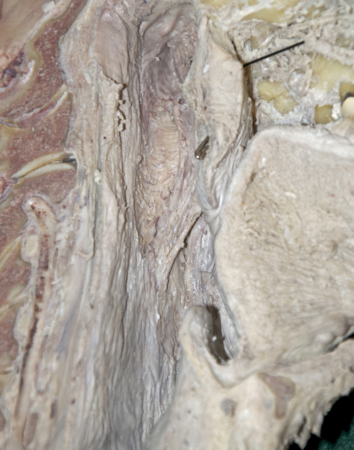
- Identify the
palatoglossal and
palatopharyngeal muscles. (G 8.32B;N 68;Gl 44.29C)
Important Relationship
- The palatoglossal fold (muscle) is positioned directly anterior to the tonsilar bed.
- The palatopharyngeal fold (muscle) is positioned directly posterior to the tonsilar fold.
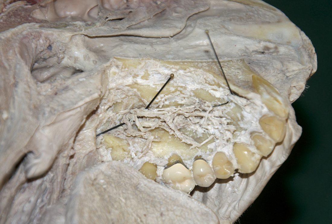
- Identify the greater palatine artery and nerve. (G 7.63D;N 57;Gl 44.16B) adjacent to the third molar where they emerge from the greater palatine foramen. Attempt to trace the greater palatine artery to the incisive fossa where it contributes to the blood supply of the anterior aspect of the nasal cavity.