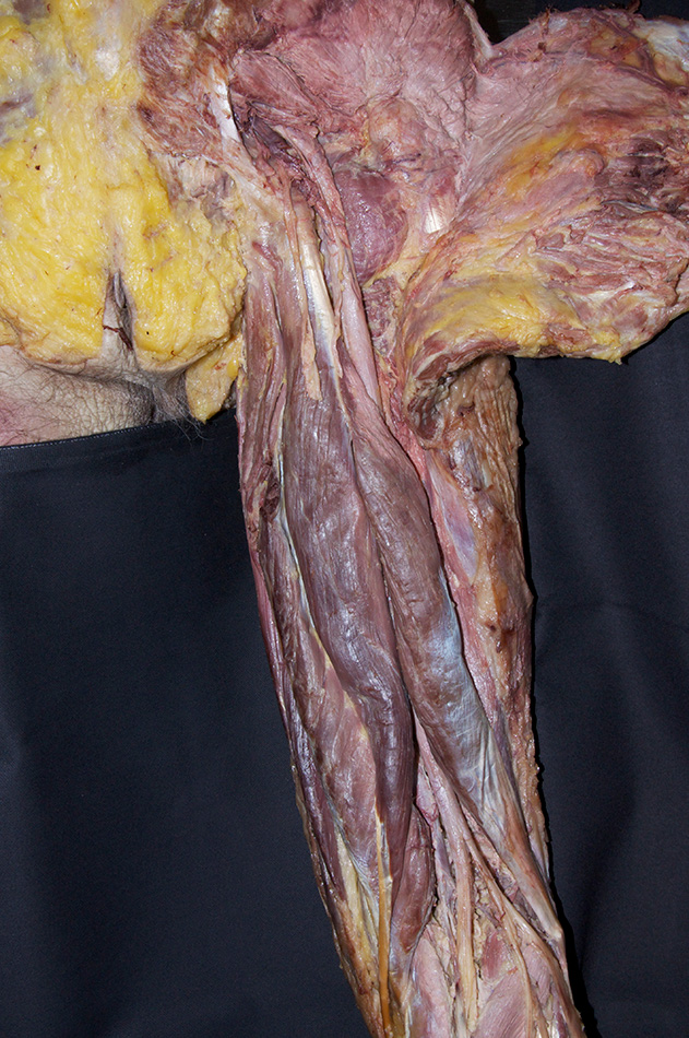
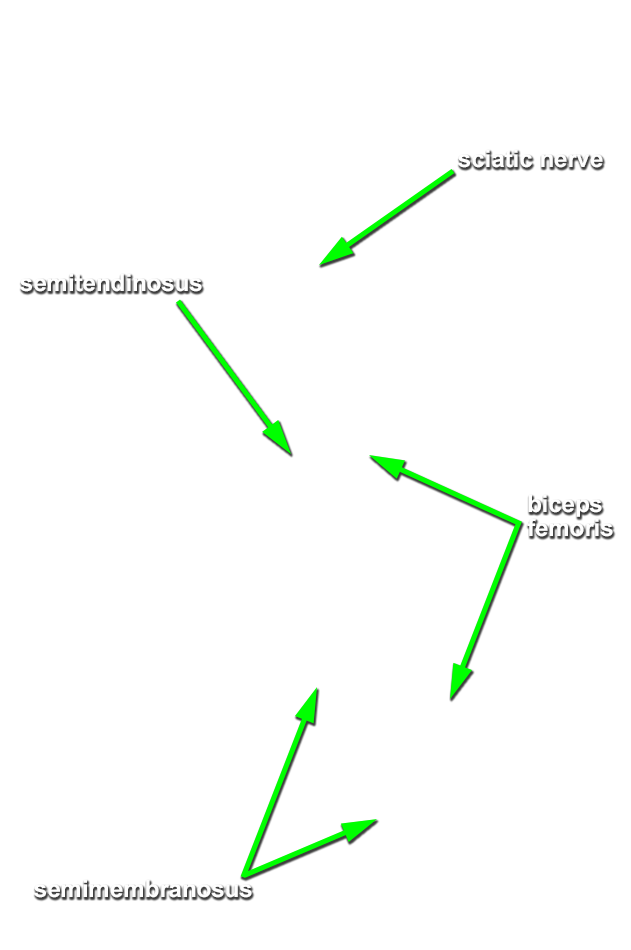
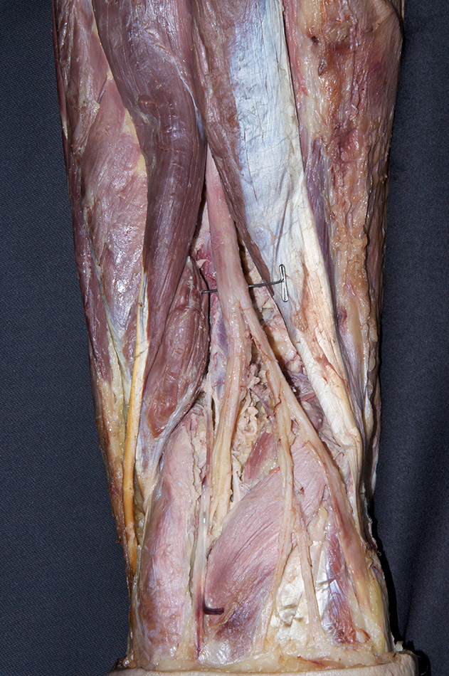
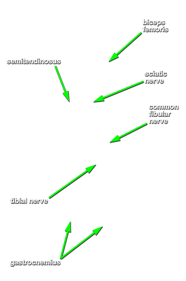
Explore the posterior compartment of the thigh. (G 6.28B;N 482;Gl 31.14B)
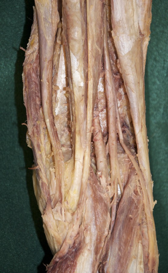
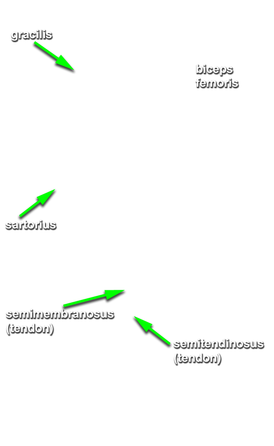
- Identify the semimembranosus and semitendinosus muscles. Trace these muscles from the ischial tuberosity to their distal attachments to the proximal, medial tibia . Note the arrangement of the distal attachments of the sartorius, gracilis and semitendinosus muscles (pes anserinus). (G 6.24A;N 480;Gl 31.13)
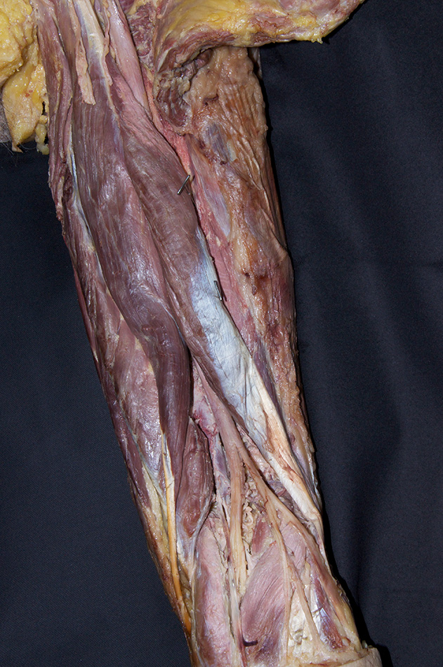
- Identify the long and short heads of the biceps femoris muscle.
- Observe the branches of the sciatic nerve entering the semimembranosus, semitendinosus and biceps femoris muscles.
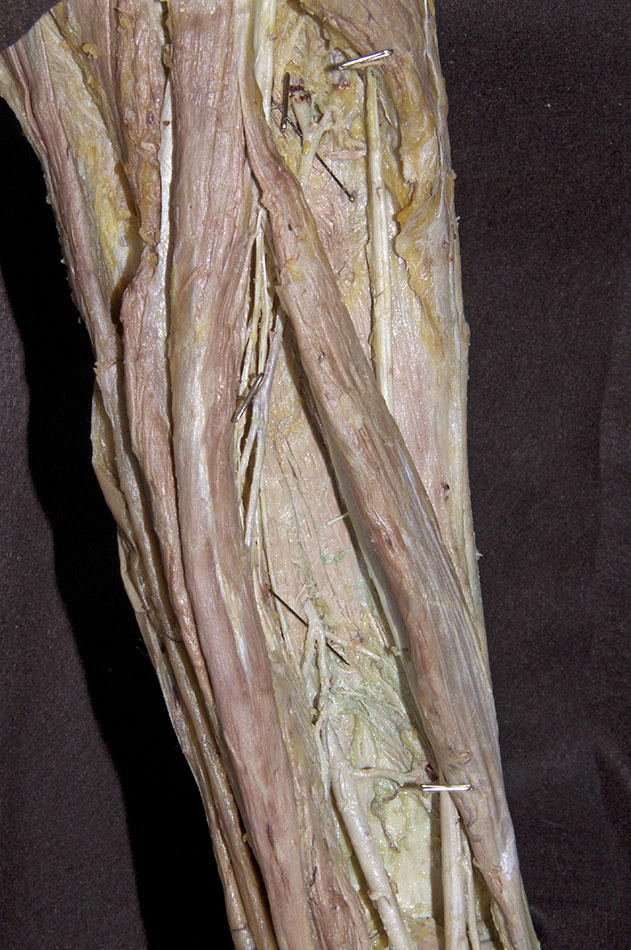
- Identify the posterior aspect of the adductor magnus muscle.
- Attempt to identify the perforating branches of the profunda femoral artery passing through the adductor magnus muscle to supply the muscles of the posterior compartment of the thigh.
- Identify and trace the sciatic nerve in the inferior direction to the point where it divides into the tibial and common peroneal (fibular) nerves. (G 6.42;N 489;Gl 34.35B)
Important Relationships
- At a mid-thigh level, the sciatic nerve is positioned posterior to the femur (shaft) and deep to the hamstring muscles.
- Within the popliteal fossa, the common peroneal (fibular) nerve is positioned lateral to the tibial nerve.